Spectrophotometric analysis, also called spectral scanning, of biomolecules serves two main purposes – the quantitation of nucleic acids (DNA and RNA) and purity assessment. Since the amount or concentration and purity of the DNA or RNA in a solution significantly affect the results of subsequent downstream applications, establishing these values early on can reduce, if not totally eliminate, the risk of committing errors and guarantee optimum results.
The Use of Spectral Scanning in Nucleic Acid Purity Assessment
Topics: Molecular Biology, Apoptosis Assays, Assay Development (ELISA), Detergents, Buffers & Chemicals, Cytology & Histology
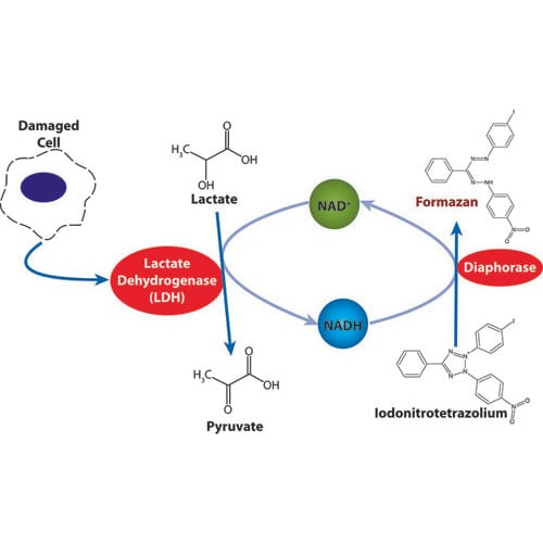
Analyzing the effects of necrosis and apoptosis is a vital component of biological research. Although there are numerous assays to detect apoptosis, there are significantly fewer assays available to measure necrosis. The release of LDH is a beneficial method for detecting necrosis, especially when combined with cellular cytotoxicity.
Topics: Apoptosis Assays, Cytotoxicity Assays
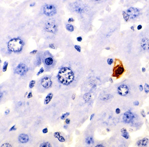
At a cellular level, there is always a homeostasis maintained between cell division and cell death. The process of growth and development involves an active balance of both of these mechanisms. Cell death occurs through three distinct mechanisms depending upon the molecular signal: Apoptosis, Necrosis, and Autophagy. All these processes have specific morphological and biochemical hallmarks that help them to get identified through different techniques. Also, these processes are crucial in a normal cellular state, while a heightened or reduced rate of these processes lead to diseased conditions. Autophagy involves intracellular protein degradation and is an important aspect of cell starvation, aging, and differentiation. Apoptosis is also known as programmed cell death, where a strict tightly controlled event making apoptotic bodies to promote cell death. Necrosis results into the release of cellular components in the extracellular space after cell destruction.
Topics: Apoptosis Assays
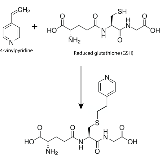
4-Vinylpyridine as a derivatizing agent for free thiols and its application in Glutathione assay
Glutathione assay involves quantification of reduced Glutathione (GSH) and Oxidized Glutathione (GSSG) in various organisms or in various tissues, blood samples, plasma, serum or cultured cells. Free thiols such as GSH can be detected by their property of relatively high reactivity compared to other biological molecules. On contrary disulfides such as GSSG does not have any unique property that could be exploited for its quantification. Hence Glutathione disulfide (GSSG) quantification can be done after reducing it to its corresponding thiol (GSH). Thus the most widely adopted methodology for quantification of GSH and GSSG is determination of total GSH concentration, followed by alkylation to remove GSH and then reduction of GSSG and its quantification. GSH concentration in sample can be determined by subtracting glutathione disulfide concentration from total glutathione concentration.
Topics: Apoptosis Assays


