 Analyzing the effects of necrosis and apoptosis is a vital component of biological research. Although there are numerous assays to detect apoptosis, there are significantly fewer assays available to measure necrosis. The release of LDH is a beneficial method for detecting necrosis, especially when combined with cellular cytotoxicity.
What is LDH?
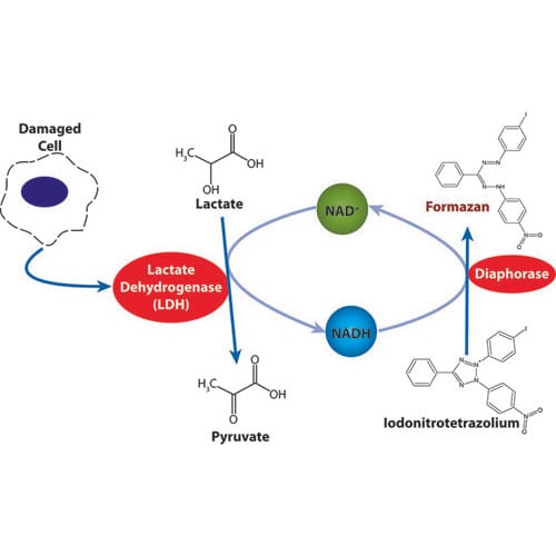
Lactate dehydrogenase, or LDH, is a stable, soluble cytosolic enzyme that is present in most eukaryotic cells. When the plasma membrane is damaged, LDH releases into the cell culture upon necrosis (accidental cell death) or apoptosis (programmed cell death).
Next, there is an enzymatic reaction that measures LDH and results in the conversion of a tetrazolium salt into a red color formazan by diaphorase.
A reliable cytotoxicity assay detects cell toxicity and cell death, as well as cell viability and cell proliferation. The assay is also ideal for the application of cell-free supernatants from cells in culture (adherent or suspension).
What is Cellular Cytotoxicity?
Cellular cytotoxicity refers to the ability of particular chemicals or mediator cells to destroy living cells. By using a cytotoxic compound, healthy living cells can undergo necrosis or apoptosis.
Also known as antibody-dependent cell-mediated cytotoxicity, cellular cytotoxicity is an immune reaction. The target cell or microbe is coated with antibodies and killed by a specific type of white blood cell. Then, the white blood cells bind to the antibodies and release substances that kill the target cells or microbes.
The ability to accurately measure cytotoxicity is valuable when identifying compounds that pose potential health risks to humans. This step is beneficial during the research phase of developing new pharmaceutical products and medicines to ensure the safety of users.
By understanding the mechanisms of cytotoxicity, researchers can gain in-depth knowledge about the standard and abnormal biological processes of governing cell growth, cell proliferation, and cell death.
How Does LDH Impact Cellular Cytotoxicity?
When a cytotoxic compound is present, living cells either undergo necrosis or apoptosis. When cells undergo necrosis, they swell and lose membrane integrity before they shut down and release their intracellular contents into the surrounding environment. Typically, external factors such as toxic chemicals or traumatic physical events trigger necrosis. However, cells undergoing apoptosis go through a series of well-defined events, such as the shrinking of the cytoplasm or cleavage of DNA into smaller fragments before white blood cells engulf them.
When the cell membranes are compromised or damaged, lactate dehydrogenase releases into the surrounding extracellular space. When lactate dehydrogenase is present in the cell culture, it reduces NAD+ to NADH and H+ through the oxidation of lactate to pyruvate.
Afterward, the catalyst (diaphorase) transfers H/H+ from NADH+ and H+ to the tetrazolium salt INT to form the red color formazan salt. The amount of color produced is measured at 490nm by standard spectroscopy and is proportional to the number of damaged cells in the culture.
Choose G-BioSciences for Your Cellular Cytoxicity Needs
At G-Biosciences, we offer a variety of products for your LDH and cellular cytotoxicity applications. To place your order, visit our website or contact us today.