
 Following up the Immunoprecipitation Protocol: The Basics, we explore the IP protocol from the viewpoint of some of the protocols critical parameters.
- Antigen extraction: As mentioned in the general protocol, cellular antigens are required to be extracted from a suitable source depending on the experimental requirement. The extracting method should be able to make antigens accessible for binding with the specific antibody. The antigens should be in a physical form in order to allow its separation from other cellular components. Hence, extraction with nondenaturing detergents such as Triton X-100, Nonidet P-40, CHAPS, digitonin, or octyl glucoside can allow the exposure of epitopes in the antigen for better binding with antibodies. Some of these detergents can even preserve weak protein-protein interactions, for example, digitonin. Some antigens are insoluble in non-denaturing detergents such as cytoskeletal structures, chromatin, membrane “rafts” or the epitope is hidden between the folded motifs of the proteins, and in that condition it is preferred to extract the antigens under denaturing conditions.
- Antigen Concentration: The cellular abundance of the immunoprecipitated antigen is essential to determine efficiency of immunoprecipitation. Radio labelling or non-radiolabelled methods are suitable to detect antigens as low as 10,000 to 100,000 copies per cell, which is generally the case with signal transduction proteins, transcription factors or endogenous integral membrane proteins.
- Pre-Clearing of the protein sample: In order to start immunoprecipitation, pre-clearing of the antigen sample is an additional step. The samples are incubated with the beads (Protein A and G or A/G) in order to get rid of unwanted antigens. The non-specific antigens get bound with the solid phase and hence can be removed by low-speed ultracentrifugation.
- Antibody purification method: Polyclonal antibodies used for IP could be in the form of whole serum, ammonium sulphate–precipitated immunoglobulin fractions, or affinity-purified immunoglobulins. Among all these, affinity-purified polyclonal antibodies are preferred as they are relatively specific and give lesser background.
- Antibody selection- Polyclonal versus monoclonal: The selection of type of antibody most suitable for IP is most important, though it is not an easy way.
| Type of Antibody | Pros | Cons |
| Polyclonal antibodies |
|
|
| Monoclonal antibodies |
|
|
Hence, an informed empirical approach should be used to choose the antibody to get satisfied results.
- Antibody titer: The titration of the antibody is required to get an optimal result. Too less of antibody will give inefficient immunoprecipitation while too much of antibody can create nonspecific IP because of increased amount of immunoglobulins bound with the beads.
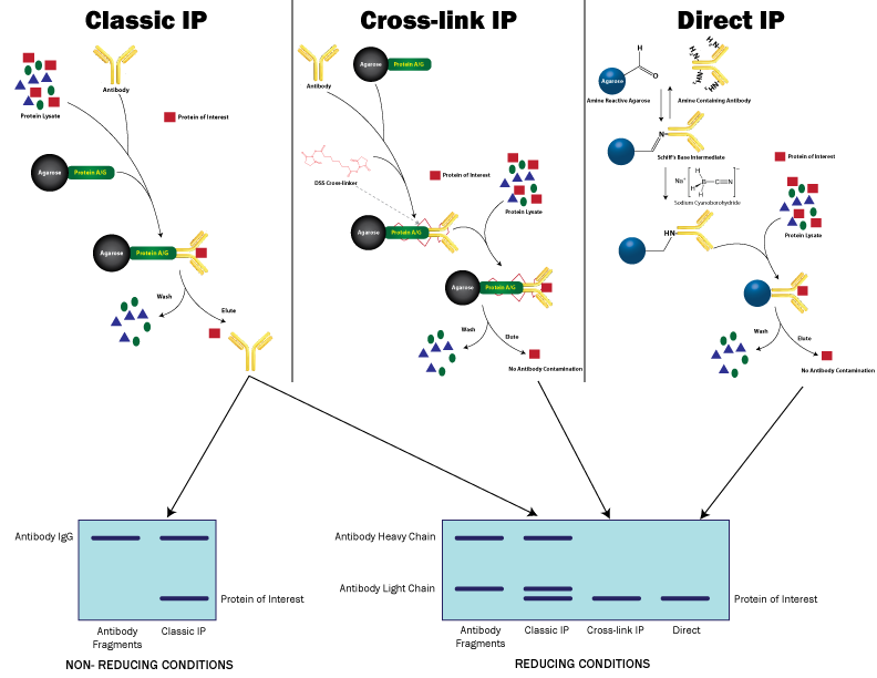
- Immunoadsorbents: Immobilized proteins (protein A, G, A/G and recombinant proteins) are efficient for antibody adsorbent and provide a beaded support. Click here for a comparison of these immobilized proteins in terms of Immunoglobulin binding.
Both Protein A and protein G are relatively stable and can be sedimented easily by low -speed centrifugation. Although, some polyclonal or monoclonal antibodies may not efficiently bind with all protein A or protein G, which can be solved by using an intermediate antibody to the immunoglobulin of interest. - Nonspecific controls: For correct interpretation of the result, it is critical to include appropriate controls for any experiment. For Immunoprecipitation, appropriate nonspecific controls with the specific samples are used. Generally two types of controls are used:
- Incubation of an irrelevant antibody similar to the experimental antibody which share biochemical similarity and belong to same species and immunoglobulin subclass. "No antibody" controls are strongly not advised.
- Perform an Immunoprecipitation from the cells/tissues that do not express a specific antigen along with an immunoprecipitation of the antigen-expressing samples.
- Washing: The washing steps must be optimized for sufficient removal of unbound proteins. Additional washes are most likely to reduce antigen-antibody association while insufficient washes may lead to background. The last PBS wash is required to remove detergent from the complex which might reduce the resolution on SDS-PAGE and other components of the buffer.
- Analysis of Immunoprecipitation result: The immunoprecipitated antigens can be analysed by different ways such as Western blotting, ELISA and mass spectrometry.


