
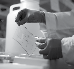
 While several protein separation technologies have been developed in recent years, two-dimensional gel electrophoresis remains one of the most popular techniques used in the field of proteomics. This is not surprising since 2D PAGE is routinely used to accurately analyze the individual components of even the most complex proteins, and can simultaneously resolve more than 5000 proteins (depending on the gel size used).
However, due to the inherent flaws in the current protein precipitation methods, 2D gels are susceptible to horizontal and vertical streaking. Here are some of the factors that may trigger this problem.
- sample preparation problems (e.g. presence of ionic contaminants such as salt, detergents, phenolic compounds, lipids and nucleic acids)
- poor protein solubilization
- high sample load (protein overloading)
- incomplete or excessive isoelectric focusing
Additionally, electroendoosmotic flow may also cause the appearance of horizontal strips. On the other hand, vertical streaking may also be brought about by any of the following factors:
- insufficient equilibration
- protein oxidation
- improper placement of the IPG strip on the second dimension gel (e.g. when the IPG strip is not in direct contact with the gel)
- uneven pore size resulting from the thinning or swelling of some sections in the IPG strip
In some cases, the 2D pattern may also be distorted due to the uneven polymerization of the gel. Vertical gaps in the 2D pattern (different from vertical streaking) may likewise be caused by excessive DTT in the sample buffer, the presence of air bubbles on the IPG strips (specifically in the agarose overlay), and/or a torn or insufficiently rehydrated IPG strip.
Improving Spot Resolution: 7 Tips to Ensure Better Results
What can you do to increase the number of spots, improve spot resolution and reduce the appearance of artifactual spots in your 2D gels? Here are some tips that can help you get better results.
- Use analytical grade quality reagents. Always use fresh solutions in your experiments. Avoid reusing your running buffer and don’t get into the habit of repeatedly freezing and thawing your solutions and samples.
- Solubilize the sample. Since repeated precipitation and resolubilization increases the risk of streaking, use the appropriate reagents to make sure all the proteins in the sample are completely solubilized prior to application. You may also centrifuge the sample solution to remove all insoluble protein complexes that may clog the pores of the gel matrix.
- Limit your contaminants. The presence of interfering agents (e.g. ionic detergents, metal ions, substrates, inhibitors, and other charged molecules) may lead to streaking, underfocusing and excessive IPG heating. Avoid these problems by using high-quality cleanup products such as Perfect-FOCUS and PAGE-Perfect to selectively precipitate your proteins from the solution. Treating the sample with nucleases can help remove nucleic acids while ultracentrifugation can remove carbohydrates and nucleic acids from the solution.
- Avoid overloading the IPG strip. The risk of horizontal and vertical streaking increases when you load large quantities of protein and/or when using samples containing highly abundant proteins. To get cleaner and more reproducible results, load less protein in the IPG strip. You can also get a sharper resolution by using a more sensitive stain with abundant proteins.
- Run samples separately. Avoid running samples with different conductivities together. The more conductive samples will naturally attract most of the current and affect the integrity of your results.
- Rehydrate IPG strips. IPG strips need to be properly rehydrated prior to use to enhance their absorption capacity and to make sure that all the proteins in the sample are uniformly distributed throughout the entire length of the strip. To ensure proper rehydration, remove all air bubbles between the IPG strip and the bottom of the tray, and don’t let the strips stick to the bottom of the tray. Incubate the strips for about 12 hours at 20oC to restore the original thickness (about 0.5 mm) and make sure they are covered with mineral oil to avoid dehydration.
- Start low. Don’t exceed the recommended voltage since it will distort your results. Better start low and gradually increase the voltage during the run.


