Proteinase K is a serine protease, the presence of a catalytic triad characterizes serine proteases, the catalytic triad is a cluster of three amino acids that make the catalytic center and consists of serine, aspartic acid, and histidine amino acids, which can often vary but all of these enzymes have a nucleophile serine and the same catalytic mechanism. Proteinase K has a catalytic triad consisting of Ser 224, His 69, and Asp 39. The substrate recognition sites are made up of peptide chains, 99-104 and 132-136.
Proteinase K cleaves peptide bonds next to the carboxyl-terminal of aromatic amino acids, hydrophobic amino acids and sulfuric amino acids, within the polypeptide chain. Hence Proteinase K can act as an endoprotease on a wide range of protein substrates. Because of its proteolytic preferences, it is a member of the subtilisin group of serine proteases. The K in Proteinase K refers to its ability to digest Keratin protein found in hairs, nails and hooves. Proteinase K is an essential reagent in molecular biology labs because it is useful in isolating DNA and RNA molecules by degrading and inactivating proteins. Fungus Tritirachium album Limber naturally produces proteinase K.
The stability of Proteinase K
Proteinase K can hydrolyse various peptide bonds under diverse conditions, even under ones that are too stringent to render other proteases inactive. For example, Proteinase K remains an active wide range of temperatures; it can remain active up to 65°C and remain active for a broad range of pH between 7.5 and 12.0. The activity of Proteinase K is somewhat enhanced in the presence of chaotropic agents such as SDS (up to 1-2%)1,2 and Urea (4 M)3. Calcium stabilizes Proteinase K for thermostability of Proteinase K, although not essential for catalysis, and hence Proteinase K remains active in the presence of EDTA (4). Proteinase K should be stored at -20°C but is also stable at 4°C.
When to use Proteinase K?
As mentioned above, Proteinase K is active in harsh conditions making it an excellent choice for use with various buffers and cell lysis conditions and allows its use in a variety of applications. Such as the preparation of chromosomal DNA for pulsed-field gel electrophoresis, protein fingerprinting5 and more commonly removal of nucleases from DNA and RNA preparations. The mechanical or detergent treatment of cells that precedes DNA or RNA isolation damages cellular ultrastructure and releases nucleases such as DNAse or RNAse that can damage DNA/RNA before isolation. Proteinase K digests all the proteins and nucleases and facilitates isolation of intact DNA/RNA molecules.
How to use Proteinase K ?
A typical working concentration for Proteinase K is 50–100μg/ml.
Proteinase K's activity has also been defined in Anson, where one Anson Unit (AnsonU) is defined as the amount of proteinase K required to liberate Folin-positive amino acids and peptides, corresponding to 1 μmol tyrosine under assay conditions in 1 minute using haemoglobin as a substrate6.
After proteinase K digestion of cellular lysates, it is crucial to perform phenol extractions to separate high molecular weight DNA. The removal of proteinase K. Phenol extraction also enables phase portioning lipids, proteins and DNA distinctly. Lipids stay in the organic phase due to hydrophobic character; the DNA resides in the aqueous phase7. Protein (including proteinase K) stays at the organic/aqueous interphase. Clean DNA is then extracted from the aqueous layer.
Proteinase K is also used for RNA and mRNA Isolation, the addition of proteinase K degrades and inactivates even traces of ribonuclease. Again, the additional phenol-chloroform and isopropanol precipitation brings down the protein contamination and makes it easier to dissolve the final pellet.
Lyophilization of Proteinase K
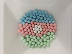
Lyophilization is a process that “freeze dries” proteins or other components. The material is frozen under reduced pressure which allows sublimation of water and works to preserve the activity and extend the shelf. Lyophilization in many cases reduces the constraints on storage temperature and metered aliquots reduce material loss. G-Biosciences has recently unveiled a convenient lyophilized proteinase K that is available in a 1mg/bead format.
https://www.gbiosciences.com/Gdotproteinasek
How to quality check Proteinase K?
The proteinase K purity can be a critical determinant for downstream applications with purified DNA/RNA, and therefore it is important to check the Proteinase K quality. This can be done using the following simple tests.
DNA Digestion test: Here 1μg of lambda-DNA and 1.8 units enzyme are incubated together for 16 hours at 37°C. For a pure Proteinase K, a same DNA band pattern after gel electrophoresis should be seen with and without Proteinase K.
RNA Digestion test: Here Incubation of 6 units of the enzyme with 1 μg MS2 RNA is carried out in assay buffer for 4 hours at 37°C. A pure Proteinase K should have the same RNA band pattern with or without Proteinase K treatment.
References:
- Bajorath, J. et al. (1988). Biochimica et Biophysica Acta. 954, 176-182.
- Ebeling, W. et al. (1974). Eur. J. Biochem. 47, 91-97.
- Hilz, H. et al. (1975). Eur. J. Biochem. 56, 103-108.
- Bajorath, J. et al. (1988). Eur. J. Biochem. 176, 441-447.
- Petrotchenko E V, et al. (2012) Mol Cell Proteomics. 11(7)
- Anson M.M, (1939) J. Gen. Physiol., 22 : 79, 1939
- Ahmad, N. N. (1995).Journal of Medical Genetics, 32(2), 129.


