
 Proteins separated by gel electrophoresis can be visualized using different staining procedures, including Coomassie stains, silver stains and fluorescent stains. Among these staining techniques, silver staining undoubtedly offers the highest sensitivity. However, it also involves a complex and relatively time-consuming protocol, has a low linear dynamic range and reproducibility, and is not compatible with mass spectrometry.
Fluorescent stains, on the other hand, offer high sensitivity and a wide linear dynamic range. It also has a simple yet robust protocol and is compatible with mass spectrometry. However, fluorescent dyes are also more expensive as compared to the other techniques and requires a couple-charged device (CCD) camera or fluorescence scanner for imaging. For these reasons, most researchers favor the Coomassie staining technique instead.
Coomassie Dyes 101: Understanding the Basics
Coomassie stains (also known as Coomassie Blue or Coomassie Brilliant Blue) are the most popular anionic protein dyes used in visualizing proteins in SDS-PAGE gels. While they may not be as sensitive as fluorescent and silver stains, these dyes provide good quantitative linearity and medium sensitivity. These qualities make it the most widely used staining technique for protein identification and mass spectrometry in protein laboratories all over the world.
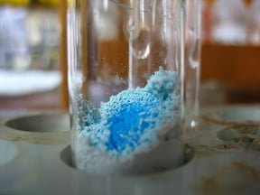
Coomassie dyes are commonly used as the basis of stains for detection of proteins in gel electrophoresis and Bradford-type assay reagents for protein quantitation. They come in two variants – Coomassie R-250 and Coomassie G-250. Basically, they are two chemical forms of a disulfonated triphenylmethane compound, and are structurally very similar, aside from the fact that G-250 has two additional methyl groups. Coomassie dyes work by binding to proteins through ionic interactions between the sulfonic acid groups in the dye and the positive amine groups in the protein, and through Van der Waals attractions.
R-250 vs. G-250: Which One Should You Use?
Basically, both R-250 (R signifies the slightly reddish tint in the blue color of the dye) and G-250 (G signifies the greenish tint in the dye) have relatively high sensitivity and allow for easy detection (they develop intensely colored complexes upon binding with protein molecules). They also allow reversible staining under the proper conditions.
Although they have basically the same chemical structure, R-250 and G-250 require different physical and staining procedures, and are not interchangeable in any particular protocol. They do not bind equally to all proteins, and do not react chemically with proteins but merely form non-covalent complexes.
Between the two, Coomassie R-250 is the more commonly used variant for protein detection since it can be used to detect as little as 0.1 ug of protein. When using the classical staining technique, the protein gels are incubated with a Coomassie staining solution and then destained using an acetic acid solution to allow for the visualization of the protein bands. This relatively cheap technique involves a simple protocol and is compatible with mass spectrometry but it has a low reproducibility and may make visualization of low abundance proteins difficult.
Like R-250, Coomassie G-250 (also known as colloidal Coomassie dye) also offers relatively high sensitivity and involves a simple protocol. However, G-250 offers a faster staining protocol and eliminates the need for destaining the gel (you can easily visualize the protein bands against the light amber background).
In addition, G-250 can be used to quantify the amount of protein in the solution (Bradford assay). Upon binding with proteins, the dye (which has a brownish color under acidic conditions) will produce a bluish tint. The optical absorbance of the solution is measured at a wavelength of 595 nm and the resulting data can then be used to determine protein concentration and the actual amount of protein in a given solution.
Image Source : Iwan Gabovitch


