
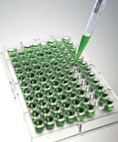
 When it comes to detecting and quantifying the concentration of an antigen in an unknown sample, most researchers depend on ELISA (Enzyme-Linked Immunosorbent Assay). This is completely understandable since this plate-based assay technique has a completely robust nature, exhibits exceptional sensitivity and is quick and easy to perform.
If this is the case, why do your ELISA experiments give less than optimal results? What should you do when this happens? Here are some of the most common problems researchers run into when performing ELISA experiments along with some of the possible solutions.
Weak or Low Signal Intensity
Are you repeatedly getting readings below the lower limit of absorbance? Does the concentration of your target analyte fall outside the range of your standard curve? If you are having these problems, you may be having issues with your reagents, your antibody, or the plate was read at the incorrect wavelength. If you think that your problems are caused by any of these factors, here are some tips to set things straight.
- Make sure reagents are at room temperature before starting the assay.
- Store reagents at 2-8oC and check expiration dates before using.
- Make sure reagents are prepared correctly and added in the proper order.
- Check reagents for interfering buffer or sample ingredients (e.g., sodium azide, EDTA).
- Always follow recommended antibody dilutions.
- Try increasing incubation times for your antibodies to ensure maximal antibody binding and amplify the signal.
- Increase the HRP concentration by 50% to increase the signal.
- Use the recommended wavelength or filter and set the plate reader accurately for the type of ELISA substrate
High Background
Getting values above 0 in the negative control wells on the ELISA plate indicates a high background signal which can ruin the results of the entire assay. This is not what you want, right? Since high background errors may be caused by problems with your antibody, buffer, substrate and/or plate, here’s what you need to do to make sure you don’t encounter this problem.
- Consider reducing your antibody concentration or run an optimization trial to determine the concentration that will deliver optimal results.
- Run negative control to determine if cross-reactivity is causing the problem.
- Prevent non-specific binding of antibodies by using affinity purified antibody and suitable blocking buffers. Pre-processing the wells according to protocol recommendations will also help.
- Store substrate in a dark place and carry out substrate incubation in the dark.
- If your buffers are ineffective or contaminated, try using a fresh buffer or a higher blocking protein concentration. You may also consider increasing blocking time.
- High background may also result from sub-optimal salt concentration in the wash buffer. If this is the case, try increasing the salt concentration to reduce non-specific interactions.
- Dirty or defective plates can cause high background so wash plates according to protocol recommendations.
High Well-to-Well Variation
If you’re getting highly inconsistent results, there is a high probability that you are committing errors in plate handling/loading or in preparing or using your reagents. Consider these tips to prevent these problems from ruining your results.
- Do not stack plates during incubation to ensure even temperature distribution and make sure plates are warmed evenly before use to avoid edge effects.
- Make sure that there are no bubbles in the wells prior to incubation and before the reading of the plate.
- Wash all wells thoroughly since uneven washing may cause large coefficient of variation (CV) and skew your results.


