
Purified and commercial antibodies are routinely stored in buffers that contain the proteins bovine serum albumin (BSA) and gelatin as these act as stabilizers during long term storage. The presence of 0.2-1% (2-10mg/ml) BSA and/or gelatin help to stabilize antibody solutions that are less than 1mg/ml during long term storage. For the majority of immunodetection techniques (ELISA, Western blotting, immunoprecipitation) the presence of the stabilizers do not interfere with these immunodetection techniques and can be used without clean up.
The presence of concentrations of stabilizer proteins, that are typically at a higher concentration than the antibody, interfere with labeling and bioconjugation techniques, such as fluorescent labeling, biotinylation and covalent immobilization of the antibody. If these techniques are used then the antibodies need to be affinity purified to remove the interfering proteins.
There are essentially three methods that can be used to remove BSA or gelatin from the antibody solutions:
- Cibrachon Blue resin
- Protein A or Protein G resin
- G-Biosciences Pearl Resin
Cibrachon Blue Resin
Otherwise known as our AlbuminOUT™, removes BSA by binding and depleting the BSA, however Cibrachon Blue has a low capacity for BSA and has been shown to be able to bind the IgG itself.
Protein A or Protein G
Both immobilized Protein A and Protein G can be used to bind the antibody and wash away the stabilizer proteins (BSA and gelatin), however the elution methods to release the antibody are typically harsh and these can affect the antibody function. In addition, once eluted the antibody will require further clean up (dialysis or desalting) to exchange the elution buffer for a suitable labeling or conjugation buffer.
Pearl Resin
 The Pearl™ IgG Purification Resin allows for the one-step purification of immunoglobulin G from serum, cell culture supernatant and ascites fluid. The resin binds the high abundant, non-IgG proteins (i.e. albumin and/or gelatin) and allows the IgG molecules to pass through in a physiological buffer. The IgG molecules can be stored or used in downstream applications without further clean-up, such as ammonium sulfate precipitation. G-Biosciences Antibody Clean Up kit is specifically designed for the rapid clean up of antibody solutions using a combination of our Pearl™ IgG Purification Resin to remove the protein stabilizers and SpinOUT™ desalting columns to ensure the antibody solutions are in an optimal buffer for clean up. The Pearl™ IgG Purification Resin binds the high abundant, non-IgG proteins (i.e. BSA and gelatin) and allows the IgG molecules to pass through in a physiological buffer.
Protocol
|
Description |
Size |
|
Caps, Screw Micro |
10 |
|
IgG Isolation Buffer |
100ml |
|
Pearl IgG Purification Resin |
1.2ml resin |
|
Spin Column, 1ml |
10 |
|
Collection Tube, 2ml |
10 |
|
SpinOUT GT-600, 3ml |
10/bag |
Resin is a 50% slurry in 5mM sodium phosphate, pH6.6 and 20% ethanol as a preservative.
Additional Items Required
- IgG samples with up to 1% BSA and gelatin
- 15ml collection tubes
Sample Preparation: Desalting
- Centrifuge the SpinOUT™ column at 1,000g for 2 minutes to compact the resin.
- Prepare the Spin-OUT™ column by removing the top and then bottom caps. Place into an appropriate collection tube.
- Mark one side of the column and ensure in all centrifugations the mark is facing outwards during centrifugation.
- Centrifuge the column at 1,000g for 2 minutes to remove the storage buffer. This compacts the resin and removes the storage buffer.
- Place the column in a new collection tube and remove the cap.
- Add 1ml IgG Isolation Buffer to the column
- Centrifuge the column at 1,000g for 2 minutes to remove the buffer.
- Repeat steps 6 and 7 three more times, ensuring the buffer is discarded after each centrifugation.
- Place the column in a new collection tube and remove the cap.
- Slowly, apply 0.5ml antibody solution to the center of the SpinOUT™
- Centrifuge the column at 1,000g for 2 minutes to collect the desalted antibody solution. Discard the column.
IgG Purification
- Ensure the IgG Isolation Buffer and the Pearl™ IgG Purification Resin are equilibrated to room temperature before starting the protocol.
- Swirl the Pearl™ IgG Purification Resin to achieve a homogenous suspension and transfer 200µl of suspension to a column using a wide bore pipette.
- Snap off the end cap from the column and retain. Place the column in a collection tube and centrifuge the spin column at 2,000-5,000xg for 1 minute. Discard the flow-through.
- Add 300µl IgG Isolation Buffer to the column.
- Briefly centrifuge (10-30 seconds) and discard the flow through. Repeat steps 4 and 5 once.
- Add 500µl desalted antibody solution (Step 10 above) to the column and seal the column with cap and end cap from step 3. Incubate for 5 minutes at room temperature with tumbling.
NOTE: For samples with >1% BSA/gelatin, adjust the volume of resin used to ensure sufficient resin is available to bind the stabilizer proteins.
- Remove the bottom, then top, cap and centrifuge the column at 2,000-5,000xg for 1 minute to collect the purified IgG.
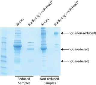
- The purified IgG is now ready for downstream applications or stored. The antibody can be evaluated by SDS-PAGE to determine the presence of BSA or gelatin


