A reliable LDH cytotoxicity assay detects cell toxicity, cell death, cell viability, and cell proliferation. It is also ideal for the application of cell-free supernatants from cells in culture (adherent or suspension).
What is an LDH Cytotoxicity Assay?
 An LDH cytotoxicity assay is a colorimetric assay that measures the stable, cytosolic lactate dehydrogenase (LDH enzyme). It is released from damaged cells and is a biomarker for cellular cytotoxicity and cytolysis.
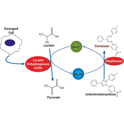
The released LDH is measured with a coupled enzymatic reaction. It results in the conversion of a tetrazolium salt (iodonitrotetrazolium (INT)) into a red formazan by diaphorase.
LDH activity is determined as NADH oxidation or INT reduction over a defined period. The resulting formazan absorbs at 492nm and can be measured quantitatively at 490nm.
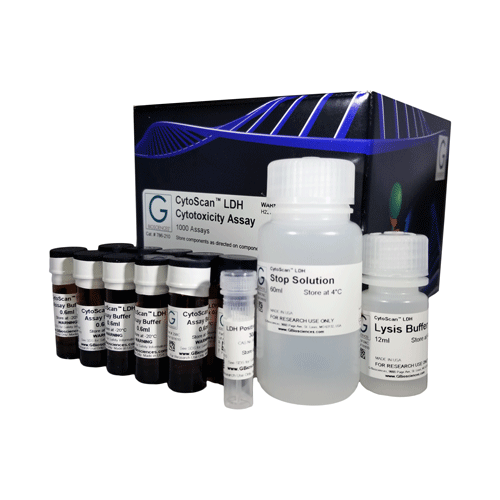
The G-Biosciences LDH Cytotoxicity Assay quantitatively measures LDH release and is ideal for high throughput screening. The kit combines reliability and simple evaluation, as well as the convenience of accuracy and avoidance of radioactivity.
The LDH Cytotoxicity Assay Kit is also compatible with different cell types for assaying cell-mediated cytotoxicity and cytotoxicity mediated by chemicals and other test compounds.
What is LDH?
Lactate dehydrogenase, or LDH, is a soluble, cytosolic enzyme found in most eukaryotic cells. LDH released into the culture medium upon necrosis (accidental cell death) or apoptosis (programmed cell death) due to damage to the plasma membrane.
Then, there is an enzymatic reaction that measures LDH and results in the conversion of a tetrazolium salt into a red formazan by diaphorase.
What is Cell Cytotoxicity?
Cell cytotoxicity is the ability of certain chemicals or mediator cells to destroy living cells. With the use of a cytotoxic compound, healthy living cells can undergo necrosis or apoptosis.
Also known as antibody-dependent cell-mediated cytotoxicity, cellular cytotoxicity is an immune reaction where the microbe or target cell is coated with antibodies and killed by a specific type of white blood cell. Next, the white blood cells bind to the antibodies and release substances that kill the target cells or microbes.
How Do You Measure Cell Cytotoxicity?
Cell cytotoxicity is measurable in several ways. However, some of the most common methods include the use of vital dyes (formazan dyes), protease biomarkers, and measuring ATP content.
The formazan dyes are chromogenic products formed by reducing tetrazolium salts by dehydrogenases, such as lDH and reductases that release during cell death. Common types of tetrazolium salts are INT, MTT, MTS, and XTT.
Likewise, cell cytotoxicity can be monitored by using the SRB assay and WST-1 assay, which are ideal for high throughput screening.
Most cytotoxicity assays work on the premise that dying cells have highly compromised cellular membranes, which enables the release of cytoplasmic components or the penetration of fluorescent dyes within the cell structure.
To eliminate the problem of underestimating cytotoxic activity because the loss of membrane integrity usually occurs quite late in the process, alternative non-radioactive bioassays measure cytotoxicity for both proliferating and non-proliferating cell samples.
What problems can occur and how can I resolve?
- High Medium Control Absorbance: There is high inherent LDH activity in the animal sera used in the culture media, try reducing the serum concentration to 1-5%.
- High Spontaneous Control Absorbance: Due to either too high of a cell density or a result of over vigorous pipetting during cell plating. Try repeating the determination of the optimum cell number for your assay and ensure that cell suspensions are handled gently.
- Low Experimental Absorbance Values: This is most likely due to a low cell density, recommend the experiment to determine optimal cell number is repeated.
- High Variability in Absorbances between wells: Check for the presence of bubbles in the wells and if present, either centrifuge the plate longer and harder or break bubbles with a syringe needle.
Rely on G-Biosciences for Your LDH Cytotoxicity Assay Needs
At G-Biosciences, we offer a selection of supplies for your cytotoxicity assay essentials. Browse our selection of products and contact us with any questions or to place an order.