
 Host cell proteins (HCPs) are one of the main impurities in bio-therapeutics that are carried over from the host expression system. Although HCPs are at low levels, they can trigger immunogenic reactions in the human system when administered along with bio-therapeutics. The similarity of physicochemical properties of HCPs and therapeutic proteins result in co-purification. Detection and elimination of these HCPs during bio-therapeutics purification are required. Development of analytical tools and methods for the detection of these HCPs during protein purification and quality control is a challenge. The final threshold level of HCPs varies for different bio-therapeutics, so manufacturers have
to analyze and report the HCP levels at all stages of downstream processing before the
product is released according to ICH-Q6B guidelines.
Different approaches to detect HCPs using methods such as Immune assays, protein assays, mass spectrometry, etc. Commercial kits are available for the detection of HCPs and they vary depending on the host organism.
Antibody-based methods: ELISA based methods are used to detect HCPs in bio-therapeutics. In this method polyclonal antibodies or monoclonal antibodies raised against host cell proteins are used. Main limitations of this assay are the inability of these antibodies to detect all HCPs of a cell type and some of them lack immunogenicity. So a successful
antibody should be able to detect proteins across wide protein concentrations from low to high. In addition to ELISA, various other methods have to be established to clearly monitor the HCPs during process development.
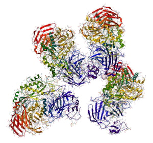
2D Gel: Various samples can be analyzed together on 1D or 2D gels in addition to other HCP detection methods. But the abundance of HCPs determines their detection though low levels of HCPs can be detected by Western blotting, it entirely depends on anti-HCP antibody pool.
Mass spectrometry-based methods: The most emerging technique useful for HCP detection is Mass spectrometry (MS). MS requires expensive equipment and skilled personnel. A large number of proteins in a sample can be analyzed in a short time by using MS. Various samples can be analyzed at a time in a high throughput manner using MS. MS analysis tools like MASCOT can be used to predict the HCP proteins. Liquid chromatography-tandem mass spectrometry (LC-MS/MS) is widely used to detect HCPs (Doneanu et al., 2012).
Presence of HCPs in therapeutic proteins cause immunogenicity and some HCPs are proteases that degrade the therapeutic protein during product development. Till date, there is no single assay that can detect all HCPs. Combination of two or more assays are used to detect different HCPs. So there is a much need to develop techniques to detect HCPs
precisely.
The acceptable limit of HCP in a therapeutic protein is 10-100 ng/mg (HCP vs Therapeutic). In case of biosimilars, in addition to the final product the amount and type of HCPs in the final sample plays an important tole in determining its similarity. So in addition to detection of these HCPs, there is much need for the availability of reference standards.
References:- Catalin Doneanu, Alex Xenopoulos, Keith Fadgen, Jim Murphy, St. John Skilton, Holly Prentice, Martha Stapels & Weibin Chen (2012) Analysis of host-cell proteins in biotherapeutic proteins by comprehensive online two-dimensional liquid
chromatography/mass spectrometry, mAbs, 4:1, 24-44. - Huang, L. Y., Dumontelle, J. L., Zolodz, M., Deora, A., Mozier, N. M., & Golding, B. (2009). Use of toll-like receptor assays to detect and identify microbial contaminants in biological products. Journal of clinical microbiology, 47(11), 3427-34.


