
Determining protein localisation and membrane topology
The subcellular localisation of protein has a major effect on its biochemical function and interaction with other proteins which in turn can have important biological implications for its function in the cell. Similarly proteins that are inserted in membranes of cellular organelles can either be facing the lumenal side of the organelle or towards the cytoplasmic side, hence the topology of proteins can vary. Very often the cell biologists are confronted by the challenge to understand the localisation and topology of a given protein. This is not an easy task and may require extensive protocols requiring extremely specific antibodies against distinct domains of the protein in question. Fortunately there is an easy way out, and this blog article deals with this simple and robust assay called Fluorescence protease protection assay or FPPA which is simple, straightforward, reliable and quick.
What is Fluorescent protease protection assay ?
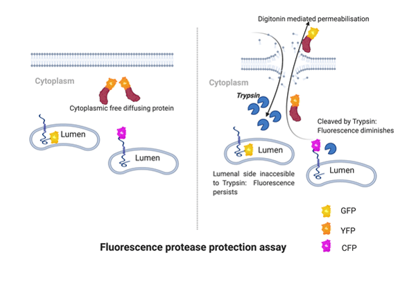
Fluorescence protease protection assay is used to detect localisation and topology of a fluorescently tagged protein. The accessibility of a proteases to exposed polypeptide versus their inaccessibility to polypeptides that are located in a protected compartment such as lumen of an organelle makes the basis of this assay. Hence the name FPPA. As mentioned earlier the protein needs to be tagged with a fluorescence marker (genetically tagged by GFP, YFP or other such fluorescent tags). The cells are then permeabilised with digitonin, digitonin is a cholesterol binding molecule derived from a plant Digitalis purpurea. Cell membranes have very high cholesterol concentration, digitonin selectively intercalates into cholesterol rich regions of the cell membranes and permeabilises but spares the intracellular membranes which tend to have a low cholesterol content. The cells are then treated with a broad spectrum protease such as trypsin. However, other approaches for cell membrane permeabilization such as streptolysin-O or Staphylococcus toxin can be used also.
How to interpret the results of FPPA?
Immediately after the digitonin mediated permeabilization, the freely diffusing cytoplasmic proteins will leach out of the permeabilised cell membrane of treated cells and hence a loss of fluorescent signal will be observed. However, if the fluorescence signal persists after permeabilization it is usually because the protein is not freely diffusing and is bound to a membrane or a subcellular structure.
Treatment of permeabilised cells with trypsin can give further information about topology of membrane proteins. To detect the topology of a given protein using FPPA, it should be tagged with a fluorophore either at N or C terminus of the protein. Even two GFP variants of the same protein expressing fluorophores at N and C terminus can be made to investigate the topology of the membrane protein. For example the termini of a type I or a type 2 membrane protein are on opposite sides, one side faces the cytoplasm and the other side is lumenal. Only the side that faces the cytoplasm is accessible to protease, whereas the lumenal side is protected from the action of proteases. Therefore the GFP variant attached to the protected site of protein will fluoresce.
Selected readings
- Lorenz H, Hailey DW, Wunder C, Lippincott-Schwartz J. The fluorescence protease protection (FPP) assay to determine protein localization and membrane topology. Nat Protoc. 2006;1(1):276-9. doi: 10.1038/nprot.2006.42. PMID: 17406244.
- White C, Nixon A, Bradbury NA. Determining Membrane Protein Topology Using Fluorescence Protease Protection (FPP). J Vis Exp. 2015 Apr 20;(98):52509. doi: 10.3791/52509. PMID: 25939013; PMCID: PMC4541577.


