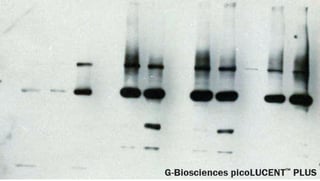
 There are a lot of reasons why researchers all over the world choose enhanced chemiluminescence (ECL) detection for a wide range of Western blot applications. ECL is considered as the experts’ detection method of choice due to its high sensitivity, excellent signal-to-noise ratio, and wide dynamic range. Additionally, ECL is also useful in quantifying a wide variety of biological materials such as cell RNA, DNA and other analytes.
Enhanced Chemiluminescence: How Does It Work?
Basically, ECL is based on antibodies that are conjugated or labeled with horseradish peroxidase (HRP). The application of a chemiluminescent substrate such as luminol or acridan and a strong oxidizing agent such as hydrogen peroxide to the blot produces excited intermediates which then release a strong blue emission at 450 nm wavelength upon their decay to a lower energy level (ground state). Since light emission only occurs during the enzyme-substrate reaction, signal output ceases when the substrate in proximity to the enzyme is exhausted.
Theoretically, the process would yield one photon of light for every molecule of the reactant used (equivalent to Avogadro’s number of photons per mole of reactant), so you can expect the light intensity to be directly proportional to the amount of the antibody bound to the target molecule. However, non-enzymatic reactions rarely do exceed 1% quantum efficiency in actual practice.
In a nutshell, the principle of enhanced chemiluminescence can best be illustrated by the following equation:
A + B→C* + D
Luminol (C8H7N3O2) + hydrogen peroxide (H2O2) → 3-aminophthalate + chemiluminescence (at 450 nm)
In a typical chemiluminescent assay, the light emitted is usually of low intensity and does not last long enough to make an accurate detection and analysis. To address this issue, most commercially available luminol is supplied with an enhancer (e.g. modified phenol, naphthol, aromatic amine or benzothiazole) so the reaction can proceed for prolonged durations without any significant reduction in light output.
By enhancing the luminol reagent, the light emission is magnified more than a thousand fold so you can accurately detect minute quantities of proteins (up to a level of 1-10 pg). In most cases, the light emission stabilizes in less than two minutes and, depending on the quantity of the reactants, may last from a few seconds to several hours (maximum of 6 to 24 hours).
The light emissions are captured with an x-ray film and/or detected by a luminescent signal instrument (e.g. charged couple device or CCD-based imager or a phosphoimager) which provides higher sensitivity, greater resolution, and wider dynamic range. Using cooled CCD cameras also allows you to do instant image manipulation and perform qualitative analysis while eliminating the need for spending time in the dark room. Despite the advantages of using digital imaging devices, it is interesting to note that most researchers still prefer to capture their data on film.
Why Choose the Enhanced Chemiluminescence Method?
The chemiluminescence method is highly recommended for the following reasons:
- High sensitivity
- Wide dynamic range
- Excellent signal-to-noise ratio
- Prolonged and intense light emissions
- Simplicity of detection
- Fast – It does not require pre-incubation steps and the substrate can be added several minutes prior to detection
- Shows a linear relationship between the concentration of the measured substance and luminous intensity
- Produces no background expression of luciferase in mammals
Some Limitations
- The light emissions produce a dynamic signal and fade exponentially over time.
- Multiple exposures are required, especially if you are using x-ray films to capture the emissions since underexposure and overexposure both produce low-quality results.
- When using x-ray films, the results may be difficult to quantify.
Further reading


