
 While researchers use several ways to visualize their protein samples, there isn’t a single best approach that is ideal for all cases. Each visualization technique has its own unique advantages and limitations, and so may only be suitable for certain applications.
Given this information, how do you know which approach to use for your particular project or experiment? Here is a rundown of the basic things you need to know about the different techniques used for in-gel protein visualization.
Dyes
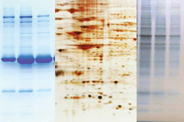
Protein laboratories around the world have traditionally used Coomassie dyes (i.e., R-250 and colloidal G-250) for visualizing protein bands resolved by SDS-PAGE. Coomassie dyes work by binding to proteins through their sulfonic acid groups via Van der Waals interactions. Aside from the two additional methyl groups in the G-250, these two variants have almost identical chemical structures.
Researchers prefer Coomassie dyes since they are simple, economical, and very easy to use. Furthermore, Coomassie dyes offer a medium to relatively high sensitivity, as they can detect proteins as low as 1 µg, and are compatible with most downstream applications (e.g., mass spectrometry, qualitative visualization, quantitative densitometry) since they do not alter the chemical structure of the target protein. And since they produce intensely colored complexes upon binding to protein molecules, they can also be easily detected in just a few minutes; maximal staining can be achieved within an hour.
Although Coomassie dyes are known to bind with a wide range of proteins, R-250 is not usually recommended if you project requires higher sensitivity. In such cases, G-250 (the colloidal form) is the better choice since it reduces the amount of free dye in the solution – which reduces background staining, eliminating the need for additional destaining steps, and enhances the sensitivity of the stain. G-250 is ideally used for quantifying the amount of protein in the solution (Bradford assay).
Ions
Silver and zinc ions are typically used for detecting proteins separated by SDS-PAGE. Silver staining protocols (i.e., silver nitrate and silver-ammonia complex methods) involve the use of simple equipment and reagents, have high sensitivity – about 10 to 100 times more sensitive than Coomassie dyes – and are compatible with downstream applications such as mass spectrometry.
Despite its desirable qualities, silver staining has its limitations. For one, it does not react uniformly with all proteins and cannot detect certain proteins (e.g., glycoproteins) due to its limited dynamic range. It also requires the use of hazardous chemicals and is temperature-dependent.
Silver ions in traditional silver staining kits may also fail to facilitate complete proteolytic digestion and maximal peptide recovery, which is a must for optimal mass spectrometry analysis. To address these limitations, G-Biosciences offers a glutaraldehyde-free silver staining kit that provides maximum sensitivity and visibility.
While silver ions cannot stain glycoproteins, phosphoproteins, and other hard-to-stain proteins, zinc ions can fill this role quite nicely. The zinc ions react with the polyacrylamide gel, turning it opaque white. The SDS prevents the stain from binding to the proteins, producing clear protein bands on the semi-opaque gel within 10 to 15 minutes. The zinc stain is also fully reversible
Fluorescent Stains
Fluorescent protein stains are versatile (can be used to stain native, denaturing, 2D, and IEF gels), highly sensitive (can detect nanogram levels of protein), have wide linear dynamic range, and offer low interference. They also provide minimal protein-to-protein variation so they can be used for quantitative protein comparison. Most fluorescent stains (e.g., Rhodamine, FITC) have simple and robust protocols, making them ideal for high throughput applications (e.g., mass spectroscopy, microsequencing, and immunostaining).
In choosing the appropriate fluorescent dye for your particular application, you need to consider its compatibility with (1) the experimental design, (2) available illumination source, and (3) other fluorescent materials in the sample which may produce background autofluorescence.


