Question:
Which non-radioactive assays are used to determine cell cytotoxicity and cell proliferation?
The Protein Man Says:
While there are a number of non-radioactive assays that can help determine cell proliferation and viability as well as cell toxicity, determining the most appropriate assay to use can be very vital in obtaining the right information from the given cell sample. So, how do you know which one to use in each particular case? Here is a guide that can help you accomplish this task.
 Measuring Cell Viability, Proliferation and Cytotoxicity
Cell viability and cytoxicity assays are best used in determining the metabolic and proliferative activities of cells within a given sample. As such, they can be used to obtain vital information on the effects of certain experimental stimulus on the proliferation of cells within an in vitro environment.
There are a number of ways by which you can get an accurate indication of the cell vitality within your sample. You can choose to assess plasma membrane integrity, mitochondrial activity and/or metabolic activity to accomplish your purpose.
Membrane integrity can be assessed by monitoring the passage of LDH to the extracellular environment. Since LDH is normally confined inside the cell, its presence in the extracellular environment indicates loss of cell membrane integrity, an occurrence that usually follows apoptosis or necrosis. If you want to measure the amount of LDH in your sample, consider using CytoScan LDH Cytotoxicity and Cytoscan Fluoro Cytotoxoicity Assays.
On the other hand, you may want to use Cytoscan WST-1 Cell Proliferation Assay if you are interested in determining cell cycle regulatory factors. This particular colorimetric assay works on the basic principle that tetrazolium salt WST-1 will be reduced to water-soluble formazan by the action of cellular dehydrogenases. The resulting formazan dye can then be accurately measured by absorbance.
For cell density determination, you can use the Cytoscan SRB Cytotoxicity Assay. This particular assay is based on the quantitative staining of cells with the fluorescent dye Sulforhodamine B, an anionic aminoxanthene dye that forms an electrostatic complex with the basic amino acid residues of proteins under moderately acid conditions. This reaction provides a sensitive linear response that can be readily measured at absorbances between 560 and 580 mm. This method is regarded to be a highly efficient and cost-effective method for screening and has a sensitivity that is comparable to those using fluorimetric methods.
In summary, here are the specific uses and properties of the different cell viability and cytoxicity assays:
CytoScan LDH Cytotoxicity Assay
-
Colorimetric assay used in measuring amount of LDH in the sample
Cytoscan Fluoro Cytotoxoicity Assay
-
Fluorometric assay used in measuring amount of LDH in the sample
-
Assays performed directly in cell culture wells
Cytoscan WST-1 Cell Proliferation Assay
-
Colorimetric assay used in determining cell cycle regulatory factors
-
Uses WST-1, a high sensitivity tetrazolium salt
-
Requires no washing, harvesting or solubilization
Cytoscan SRB Cytotoxicity Assay
-
For cell density (biomass) determination
-
Linear response
-
Simple, accurate and reproducible assay
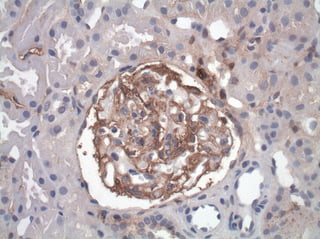
Image By: Libertas Academica


