Question
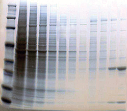
I am looking for a stain to use on a Tris-Tricine polyacrylamide gel that will stain a small peptide (~3kDa) that has no positively-charged amino acids. It's a very acidic peptide consisting only of neutral and negatively-charged amino acids. Coomassie does not work. Thank you.
The Protein Man Says:
There are a large selection of stains for detecting proteins and peptides following protein electrophoresis, however all these stains are a function of the amino acid composition of the protein or peptide. The longer the amino acid chain, the easier it is to stain, so short peptides are particularly troublesome when it comes to protein gel stains, but not impossible to stain.
There are a range of protein staining techniques and to aid in selection the correct stain The Protein Man will review the most commonly used:
- Coomassie
- Silver
- Negative
- Fluorescent
Coomassie Stains
Why Coomassie? The Coomassie dyes were originally names to commemorate the British victory over the Ashanti capital of Kumasi or 'Coomassie'; which is now in Ghana. (Fox, M.R. (1987), Dye-makers of Great Britain 1856-1976 : a history of chemists, companies, products and changes, Manchester: Imperial Chemical Industries)
The Coomassie based stains are organic stains and offer probably the simplest protein staining technique, although not the most sensitive. The Coomassie dye interacts electrostatically with charged amino acids and the Coomassie stain appears to interact more strongly with basic residues, such as lysine, histidine and arginine.
Silver Stains

Silver staining was introduced by Kerényi and Gallyas in 1973 and offers ~2,000 fold more sensitivity compared to Coomassie staining. There are several different methods of silver staining, but they all work on the principle of reducing ionic silver to metallic silver. The proteins in the polyacrylamide gel bind silver ions and these are then reduced to metallic silver that are readily visible. As with Coomassie staining, the silver ions interact more strongly with basic amino acids, particularly histidines and lysine, and the strength of staining is protein dependent.
Negative Stains

Negative or reverse stains work on the principle of forming insoluble metal salts in the gel background, leaving the protein bands or spots unstained. These stains are rapid and the unstained proteins are excellent for easy recovery and further downstream applications.
Commonly used reversible protein gel stains involve copper or zinc salts. The insoluble metal ions are repelled by the high concentration of SDS in the protein bands and are deposited in the surrounding gel matrix, leaving the clear areas or proteins. The negative stains are less dependent on the amino acid composition of the proteins and therefore has a greater ability to stain small peptides lacking charged amino acids.
Fluorescent Stains
The post electrophoretic stains, developed by Steinberg et al, used in today's research market work in a similar manner as the negative stains, in the fact that they interact with the SDS:protein complexes. The fluorescent stains bind to areas with higher SDS concentration, as opposed to the negative stains that move to areas of lower SDS concentrations.
Other Detection Approaches
If one of the above staining methods is not suitable for detecting acidic peptides then a different approach will have to be used. Several possibilities remain, including:
- Western blot transfer & staining
- Western blot transfer & antibody detection
- Pre-electrophoresis labeling
Western Blot Transfer
Following protein electrophoresis the proteins can be transfered to a PVDF or nitrocellulose membrane. Once transfered to the membrane the proteins can be detected with a membrane stain, such as Ponceau S or Amido black.
An alternative membrane stain is G-Biosciences Swift Membrane Stain that is a 30 second stain offering greater sensitivity than Ponceau-S.
If the protein or peptide of interest has a specific antibody raised against it then a classic Western blot procedure can be used to detect the protein or peptide.
Pre-Electrophoresis Labeling
A final solution for detecting proteins or peptides would be to label the protein/peptide of interest prior to electrophoresis. There are a large selection of labeling methods available to protein researchers including fluorescent tags, biotinylation and recombinant protein tags.
Download a free copy of G-Biosciences' Protein Labeling Handbook for more details.
The Protein Man Review
Staining of acidic peptides is difficult, but possible solutions are available. The first thing to try would be the reversible stains (Reversible Zinc Stain). If this fails to detect the protein then try a Western transfer and our new Swift Membrane Stain.
If all else fails then pre-electrophoresis labeling could be the only option left.
Click to download our Protein Electrophoresis Handbook
for a review of G-Biosciences protein gel stains
Do you have any tips or tricks for detecting peptides on protein gels?


