
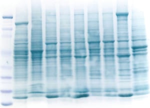
 Protein samples are analyzed by denaturing sodium dodecyl sulphate polyacrylamide gel electrophoresis (SDS-PAGE). Total protein lysates or protein samples from purification experiments are separated on 1D and 2D gels depending on their molecular weights and PI. Appropriate staining techniques are used to detect proteins on SDS-PAGE gels depending on the protein of interest, downstream experiments like Western blotting, mass spec analysis etc.,
Staining methods like Coomassie blue staining, Silver staining, Zinc staining, Fluorescent staining, Glycoprotein staining and Heme protein staining are widely used. These staining methods can be used for both 1D and 2D gels. If Western blotting is to be used then reversible stains, such as zinc should be used, or reversible membrane stains, such as Ponceau stain or commercially available reversible stains, are used to analyze the total proteins transferred to the membrane. Reversible stains need to be used for membrane staining to ensure they do not effect subsequent antibody probes. .
Coomassie blue staining: Coomassie blue staining is the widely used method for staining SDS-PAGE gels. The sensitivity of this method depends on the staining reagent used. Coomassie G-250 can detect proteins at 30 ng and Coomassie R-250 can detect proteins at 100 ng.
Silver staining: Silver staining method is one of the best methods to detect low expression proteins in a sample. It can detect proteins as low as 5-10 ng. Several variations of this method are developed but the basic steps are 1. Fixing the proteins and removing interfering compounds. 2. Sensitization to increase sensitivity 3. Infusion of gel with silver nitrate solution or Silver-ammonia complex. 4. Development of silver-protein image and 5. Endpoint development of the reaction and removal of excess silver and other impurities. Silver staining has one drawback as it doesn't stain all proteins, particularly glycoproteins and phosphoproteins.
Glycoprotein staining: Highly glycosylated proteoglycans and glycoproteins are not easily detected by Coomassie staining or silver staining due to the interference by the carbohydrate side chains. So these proteins are detected by using Periodic acid-Schiff stain.
Heme protein staining: 3,3’,5,5’-tetramethylbenzidine/H2O2 (TMBH) is used to detect heme-containing proteins like Cytochrome-C. This method is light sensitive and should be done in dark. 2-mercaptoethanol and other reducing agents interfere with the staining reaction so they should be avoided during sample processing.
Stain-free method: Roberts et al. reported a stain-free method to detect proteins within 5 minutes after running SDS-PAGE. In this method, 2,2,2-Trichloroethanol (TCE) is added to the resolving gel that later binds to the aromatic amino acids of protein samples and the proteins fluoresce under UV light. Though the method cannot detect proteins lacking Tryptophan, it can detect all other proteins. The sensitivity is more than that of coomassie staining for some proteins as reported. This method can be used for checking the gels for Western blotting as well. This is a quick method that is very useful for analyzing samples at different stages of protein purification. This is a sensitive method that can detect as low as 0.2 -0.5 ng of protein. This reagent does not interfere with western blotting and mass spec analysis.
References:
- Chevallet M, Luche S, Rabilloud T. Silver staining of proteins in polyacrylamide gels. Nature Protocols. 2006;1(4):1852-8.
- Carol L Ladner, Jing Yang, Raymond J Turner, Robert A Edwards. Visible fluorescent detection of proteins in polyacrylamide gels without staining, Analytical Biochemistry, Volume 326, Issue 1, 2004, Pages 13-20.
- B. Rivero-Gutiérrez, A. Anzola, O. Martínez-Augustin, F. Sánchez de Medina, Stain-free detection as loading control alternative to Ponceau and housekeeping protein immunodetection in Western blotting, Analytical Biochemistry, Volume 467, 2014, Pages 1-3


