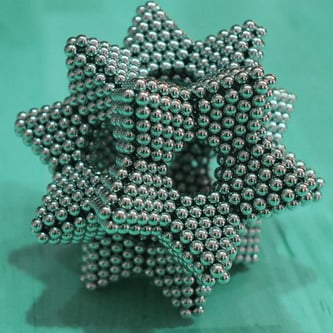
 While agarose beads have been traditionally used for immunoprecipitation (IP) and other micro-scale protein and antibody purification procedures, magnetic beads have since taken lead and are now considered tool for protein immunoprecipitation.
Magnetic Beads 101: What You Need to Know
Basically, magnetic beads are solid spherical particles around an iron core used for immobilizing molecules (e.g. proteins, enzymes, peptide, antibodies, nucleic acids) on a solid phase for the purpose of separating them from the lysate.
When using magnetic beads, all you need to do is vortex the beads before adding them to prepared sample, incubate to facilitate binding between the beads and the molecule of interest, remove unbound material through aspiration and capture the analyte-bound beads with a magnet, and elute the analyte from the beads for subsequent use in downstream applications. In some cases, however, the analyte can be used in downstream applications even while they remain attached to the beads.
Magnetic Beads: What Makes It the Better Choice?
Why do researchers choose magnetic beads for IP? Here are some good reasons why.
Magnetic beads provide optimal binding capacity. Unlike agarose beads, magnetic beads do not have a porous center that can help enhance their binding capacity. However, their relatively small size (magnetic beads have a diameter of 1 to 4μm compared to agarose beads which have a diameter of 45 to 165µm) helps them achieve optimum antibody binding capacity.
It ensures more efficient antibody consumption. Magnetic beads don’t require huge amounts of antibody to produce accurate results. They are non-porous so you can be sure that the antibody will bind exclusively on the exposed outer surface of the beads.
With agarose beads, it is an entirely different story. Their extremely porous nature increases the risk that the antibodies will get trapped inside their sponge-like structure, making them inaccessible to the target proteins. Moreover, you need to ensure that the amount of antibody used is enough to coat the entire bead or you’ll increase the risk of getting elevated background signal due to the nonspecific binding of the lysate components to the unsaturated portions of the beads.
It does not entail any sample loss. Magnetic beads do not require centrifugation, so you don’t have to worry about losing any of the bound materials (immune complexes) or breaking all those weak antibody-antigen bonds during the gentle wash steps. Thus, you can be sure you’ll get more accurate results and ensure the reproducibility of your experiment.
It is fast, easy and more convenient to use. Most researchers find magnetic beads easier and more convenient to use. Here’s why.
- Magnetic beads are non-porous, so you do not need to perform any incubation steps.
- They do not require any pre-clearing steps since there is a lower risk of non-specific binding of unwanted molecules.
- They do not need any centrifugation steps since the buffer can be removed without ever touching the beads.
When using magnetic beads for IP, you can complete the entire procedure in about 30 minutes. On the other hand, it will take you anywhere between one and 1.5 hours if you are using agarose beads.
It can be more cost-effective. Magnetic beads may be considerably more expensive than agarose beads but considering the fact that you need a significantly smaller amount in your experiment, using them can ultimately be more economical.
Ideal Applications
Magnetic beads are ideally suited for immunoprecipitation and are perfect for applications that require sample size of less than 2 ml. As such, they are commonly used for manual and automated standard IP, co-immunoprecipitation (Co-IP), chromatin immunoprecipitation (ChIP), ChIP sequencing, and pull-down reactions for immediate assay analysis.


