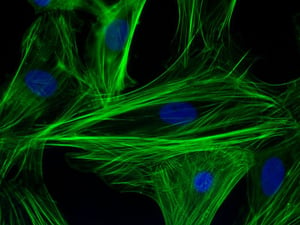
 Immunofluorescence is an essential and a powerful tool in molecular biology for the visualization of cellular structures, protein localization and intracellular processes. The resultant sample produced after immunofluorescence is analyzed using different microscopic methods such as confocal microscopy, epifluorescence, TIRF, or GSDIM depending on the experiment.
The cardinal idea of Immunofluorescence revolves around two basic components:
- Antibody specificity to mark different subcellular structures or proteins
- Type of fluorochromes used for coupling with the immune complexes for visualization of the targeted structures.
These two crucial steps determine the reliability of the results of Immunofluorescence. However, the quality of the results depends on the preparation of the biological sample. Fixation and permeabilization are the key steps that determine the success of an Immunofluorescence experiment.
FIXATION:
Fixation is the chemical preservation of the tissue for analysis by preventing cellular destruction mediated by proteases. An ideal fixative should preserve the cellular structures rapidly and uniformly. It crosslinks with the endogenous degradative enzymes and inhibit autolysis. An ideal fixative must perform following function in immunofluorescence:
- Preservation and stabilization of the tissue/ cellular architecture
- Strengthening of the tissue for further processing
- Inactivation of proteolytic enzymes to snap-shot the cellular entities
- Protection of the sample from microbial contamination and decomposition.
The method of fixation, duration and type of fixative affects all these functions and tissue integrity, therefore these fixing conditions should be optimized for an experiment.
There are two main types of fixative reagents:
- Chemical crosslinkers: They crosslink with the cellular proteins via their free amino acids and preserve the cellular morphology. These fixatives can cross the plasma membrane of the cell and crosslinks with the proteins. It allows stabilization and hardening of the tissue. The lacuna in this fixation is that these fixatives also crosslinks with the antigen of interest which might reduce signal intensity and antibody binding. Chemical crosslinkers are mostly aldehydic derivatives.
- Organic solvents: Organic solvents are dehydrogenating agents and they help in fixation by precipitating the proteins. While using these fixatives, separate permeabilization step is not required. However, these fixatives rinse off many small and soluble cellular molecules, rendering them unfit for certain experimental conditions. They also denature genetically encoded fluorescent proteins, therefore should be selected carefully.
|
Fixative Type |
Fixative |
Mechanism of fixation |
Pros |
Cons |
|
Chemical Cross linkers |
Formaldehyde or Paraformaldehyde (2-4%) |
Crosslinking with the free amino acids of the proteins |
Good for cellular preservation Ideal for genetically encoded fluorescent proteins. |
Antigen crosslinking Autofluorescence Dampening signal |
|
Glutaraldehyde |
Crosslinking with the free amino acids of the proteins |
Good for cellular preservation Ideal for genetically encoded fluorescent proteins. |
Antigen crosslinking Autofluorescence
|
|
|
Organic solvents |
Methanol (Ice-cold) |
Precipitation of the proteins |
Rapid preservation of the cells Permeabilize the cells as well |
Negatively affect protein epitopes Not appropriate for fluorescent proteins Loss of soluble and lipid molecules |
|
Acetone |
Precipitation of the proteins |
Faster preservation Does not lose many epitopes |
Not appropriate for fluorescent proteins Loss of soluble and lipid molecules |
|
|
Microwave fixation |
|
Heat induced denaturation of proteolytic enzymes |
High penetrance in thick tissues |
Loss of morphology at very high temperature |
Table 1: Comparison of different fixatives
There are two methods of fixation commonly used:
|
Type of fixation method |
Mechanism |
Suitable for |
Pros |
Cons |
|
Trans-cardial fixation (whole-body fixation) |
4% ice-cold paraformaldehyde perfused in the whole body of the animal through the vascular system |
Whole, intact tissue |
Uniform and rapid fixation |
Require technical expertise in animal handling |
|
Immersion fixation |
Tissue immersed in 4% ice-cold paraformaldehyde for overnight or longer |
small chunks of tissue |
Technically simple and requires in less time |
Uneven penetration of fixative |
Few things to be kept in mind while fixation:
- Fixation is recommended immediately after harvesting the tissue to avoid post-mortal changes.
- Fixation should be done at room temperature. High temperature may induce heat-mediated tissue damage.
- The volume of the fixative should be double the volume of the tissue at least.
- It is always recommended an overnight fixation at low temperature. Underfixation or overfixation may lead to improper staining.
PERMEABILIZATION:
Permeabilization allows the antibodies to penetrate the lipid membranes of the cell and access the intracellular structures. It is not required if organic solvents are used for fixation. For samples fixed with chemical crosslinkers require additional permeabilization step using classical detergents like Triton X-100 or NP-40 or modified detergents like saponin or digitonin depending on the antibodies, time of incubation and thickness of the sample.
Permeabilization is typically done with 0.1% detergent in PBS 15–20 minutes at room temperature. In case of lipid-associated or membrane proteins, saponin is a better detergent for the sample preparation as it selectively removes cholesterol from the cell membrane, while the intracellular membranes remain largely unaffected. Nucleus marking stains such as DAPI or Hoechst or lipid-soluble dyes like DiI can penetrate the membrane easily; therefore do not require additional permeabilization.
In order to prepare samples for final staining steps, both fixation and permeabilization should be carried optimally. If not carried properly, they might lead to improper staining:
- Underfixation: It causes slow degradation of sample and weak tissue.
- Overfixation: It leads to auto-fluorescence and masking of antigen epitopes.
- Over-permeabilization: The tissue might lose its integrity and some soluble molecules.
- Under-permeabilization: It leads to less penetrance of the antibodies inside the cell, therefore, lesser staining signal.


