
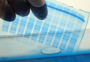
 Tris-Tricine SDS-PAGE (polyacrylamide gel electrophoresis) is used to separate protein / peptides ranging from 1-100 kDa molecular weights. This method varies from Laemmli SDS-PAGE by replacing Glycine pK (9.6) with Tricine (pK 8.15). Due to the variation in pK the resolution of high or low molecular weight proteins by both methods vary. Lower acrylamide concentrations of Tricine gels help in easy transfer of hydrophobic proteins during Western blotting. Tricine gels are suitable in isolating hydrophobic proteins from 2D gel for mass spectrophotometric analysis. It is also helpful to isolate membrane protein complexes from biological membranes. When urea is added in stacking gel it can easily separate two different proteins of the same molecular weight. Laemmli SDS-PAGE gel can separate high molecular weight proteins (20-200 kDa) but proteins less than 20 kDa are not clearly separated and diffused even if higher acrylamide concentration (4-20% gradient gels) are used. In contrast, Tricine SDS-PAGE gels can be used to separate proteins below 100 kDa only and especially 20 kDa or lower molecular weight proteins or peptides are very well separated by this method. Staining of Tricine gels is crucial as there is every chance of losing low molecular weight proteins from the gel during staining and destaining process. Fixing, Staining and Destaining should be rapid to avoid blotting out of these small proteins.
How to Prepare A Tricine SDS-PAGE Gel:
- Gel buffer: 3M Tris, 3% SDS pH adjusted to 8.45 with HCl*
- Acrylamide solution A— 49.5 %T, 3% C mixture (48g Acrylamide + 1.5 g N,N- Methylene bisacrylamide per 100ml)
- Acrylamide solution B—49.5 %T, 6% C mixture (46.5g Acrylamide + 3.0 g N,N- Methylene bisacrylamide per 100ml)
- 1X Cathode buffer (Upper tank buffer added to wells): 0.1M Tris, 1M Tricine, 0.1% SDS pH 8.25
- 1X Anode buffer (Lower tank buffer): 0.2M Tris pH adjusted to 9.0
*Note: Always adjust pH before adding SDS during buffer preparation. - 100 % Glycerol
- 10 % APS
- TEMED
- 1X Gel loading buffer (reducing) - 50 mM Tris 8 pH, 12% glycerol, 4% SDS, 0.01% Coomassie blue G-250, 2% 2-Mercaptoethanol
- 1X Gel loading buffer (non-reducing) - 50 mM Tris 8 pH, 12% glycerol, 4% SDS, 0.01% Coomassie blue G-250.
Note Coomassie blue G-250 works as best gel tracker than Bromophenol blue as it runs before the small peptides of 1-2kDa.
Separating, spacer and stacking gel composition (Adapted from Hermann etal.,1987)
|
|
Stacking gel |
Spacer gel |
Separating |
Separating |
Separating |
Separating |
|
4% A |
10% A |
gel 10% A |
gel. 16.5% A |
gel. 16.5% B |
gel 16.5% B + 6M Urea |
|
|
Solution A |
1 ml |
6.1 ml |
6.1 ml |
10 ml |
— |
— |
|
Solution B |
— |
— |
— |
— |
10 ml |
10 ml |
|
Gel Buffer |
3.1 ml |
10 ml |
10 ml |
10 ml |
10 ml |
10 ml |
|
Glycerol |
— |
— |
4 g |
4 g |
4 g |
— |
|
Urea |
— |
— |
— |
— |
— |
10.8g |
|
Total volume make up with H2O |
12.5 ml |
30 ml |
30 ml |
30 ml |
30 ml |
30 ml |
How to run a Tricine SDS PAGE gel
Based on protein or peptide need to be separated, percentage of gel is chosen. 10% gel for proteins >10kDa, 16.5% gel for peptides <10 kDa and 16.5% with urea for hydrophobic proteins. Use of both Glycerol and Urea results in sharp bands and decrease hydrophobicity of peptides. All ingredients were mixed well as mentioned in the table above and gels were cast. Either long slab gels (22x15.5x0.07 cm) or short gels (10x14x0.07cm) or 14x14 cm gels or mini gels are used depending on the purpose of the experiment. Gel loading buffer was added to the samples and heated at 40 ℃ for 30 min or 90 ℃ for 5 min and loaded in each well. Avoid over loading the protein sample, each protein band per well should be 0.5-2 µg. Tricine gel should be run at low voltage for initial 1hr (30 V) and then voltage can be increased to 180 V and gel must be kept cool by keeping the apparatus in a cold room or using precooled buffers.
Haider etal., 2010, reported a modified Tricine gel protocol for analysis of phosphoproteins by Inductively Coupled Plasma Mass Spectrometry (ICP-MS). They used 7-10 % gel to separate phosphoproteins in this modified protocol. Here they used a single buffer for running the gel (25mM Tris, 25mM Tricine and 0.05% SDS) instead of cathode and anode buffers and used 2.5 M Tris at pH 8.8 instead of 3M Tris pH 8.45 in gel buffer. They observed better resolution of small phosphorylated proteins. But many researchers follow the original protocol developed by Hermann etal., 1987.
The main drawback of Tricine gel is its running time, it takes 4-16 h. It cannot separate high molecular weight proteins. Though smaller proteins are well separated, care should be taken to avoid blotting out of small peptides during staining and electroblotting.
Apart from these drawbacks, Tricine SDS-PAGE is one of the successful electrophoresis technique used to separate and analyse small proteins and peptides by 2D gel electrophoresis, electroblotting , mass spectrometric analysis, etc.,
References:
- Hermann Schägger, Gebhard von Tricine-sodium dodecyl sulfate polyacrylamide gel electrophoresis for the separation of proteins in the range from 1 to 100 kDa, Analytical Biochemistry, Volume 166, Issue 2, 1987, Pages 368-379.
- Haider, R., Reid, H.J. & Sharp, B.L. Modification of tricine-SDS-PAGE for online and offline analysis of phosphoproteins by ICP-MS. Bioanal Chem (2010) 397: 655. https://doi.org/10.1007/s00216-010-3588-9


