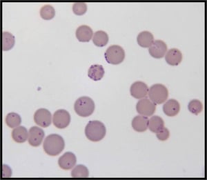
 Cell culture is extensively growing technique applied in various fields of basic research, regenerative medicines and biotechnological applications. The use of cells is exponentially increasing in wide-range of studies involving cell division, cell growth and proliferation, bioassay development and drug treatments. However, the credibility of results depends on the reproducibility of results and quality of cell culture.
Apart from general protocol factors, the quality of cell cultures relies on the purity of the culture environment. Cells are susceptible to contamination by different microorganisms as cell cultures do not have innate immune responses unlike in-vivo systems. Bacterial and fungal contaminations are most common and obvious contaminants as the detection of these is very easy and visibly observable.
The most disastrous contaminants are mycoplasma, which poses a significant alteration of cellular parameters resulting into irregularities of experimental plans and interpretation.
Mycoplasma cells are very small bacteria (less than 1 μm) without containing a cell wall, therefore extremely difficult to visually perceive using a visible light microscope. Also, unlike bacterial and fungal contaminants, mycoplasma contamination does not cause a substantial and observable change in the pH and turbidity of media making detection even tougher.
Sources of Mycoplasma contamination
Mycoplasmas grow favourably in culture supernatants and on cell membranes. Since, they are not affected by the commonly used antibiotics in the media; it provides a harvesting ground for mycoplasma growth. Different strains of mycoplasmas can contaminate cells through different resources:
- Incoming infected cells from other labs through exchange of cell vials
- M. orale, M. fermentans, M. salivarium and/or M. hominis from lab personnel
- Cell culture reagents such as bovine sera carrying M. arginine and A. laidlawii or porcine trypsin carrying M. hyorhinis.
As the cell culture system gets exposed to mycoplasma contamination, it can easily propagate through aerosol droplet dispersion. Therefore, it is essential to regularly decontaminate and fumigate the culture rooms and instruments.
How to detect Mycoplasma contaminations
Because of the small size and lack of a cell wall, mycoplasmas are largely invisible under a light field microscope; therefore require time consuming and sensitive methods to detect them. The most common and cost-effective methods for the detection are:
- Culture testing on agar, liquid media, or semi-solid media.
- Staining of the culture media with DNA stains such as DAPI Staining (4’, 6-diamine-2-phenylindole dihydrochloride).
- DNA hybridisation
- Use of antibodies specific for mycoplasmas.
- PCR based detection using specific primers.
- Enzyme-linked immunosorbent assays (ELISA), and immunoblotting.
The preferred methods are using culture based methods or PCR-based methods, which have their fair share of sensitivity and specificity.
Cell-culture based detection
- Takes time
- It takes technical expertise and it's a cumbersome protocol
- Average sensitivity
- Cannot detect all the strains of mycoplasma such as M. hyorhinis
PCR-based detection
- Fast method
- Single step process and requires common lab reagents
- High sensitivity
- Can detect all the strains
What to do if the cells are tested positive for mycoplasma contamination
- Separate the contaminated cultures from the healthy ones. Treat the contaminated flasks with the appropriate antibiotics and try to salvage the contaminated cells if possible otherwise discard them.
- Add Plasmocin to the culture media at 25 μg/mL for 1-2 weeks to eradicate mycoplasma.
- After the treatment duration, cells should be tested again for mycoplasma contamination before taken in use.
It is always advised to stay vigilant and follow the aseptic lab practices for good results.
Prevention of Mycoplasma Contamination
As it is always suggested to prevent the problem before it starts massively affecting your experiment, therefore safety measures should be taken while practicing cell culture.
- It is advised to keep a periodic scheduling of mycoplasma testing when starting a new batch of culture or freezing a stock of cell lines.
- Always follow good lab practices and wear personal protective equipment like gloves, head cover and a clean lab coat.
- Keep the lab condition sterile and aseptic by:
- Unrestricting air flow of the hood and leave it uncluttered.
- Spray the lab personnel hands with 70% ethanol before using them in the hood.
- Media bottles and plates should be kept in the hood and covered properly if moved outside
- Any spills should be removed immediately and sprayed with alcohol.
- Incubator should be cleaned periodically with the bleach and water should be changed regularly.
- In case of any doubt, quarantine the substance of doubt. Don't use it for further experiments.


