
 Poor protein transfer can lead to either a weak signal or a lack of signal being observed during the process of western blotting. Though poor protein transfer does occur fairly often, it's also commonly disregarded as a potential cause for a failure. Consequently, it's an issue that should be considered first when trying to determine the cause of a poor signal.
The Causes of Poor Protein Transfer
Poor protein transfer may occur due to low levels of antigen. This commonly occurs because of either a poor transfer procedure or a gel that was not sufficiently loaded. In a laboratory, this can occur as a mistake during the blotting process and can lead to a lack of signal. The membrane will need higher levels of antigen in order to complete the transfer successfully.
There may also be issues with the membranes themselves or with processing times, as protein transfers may take longer depending on the products used. If a protein transfer is interrupted or isn’t given the necessary amount of time, the transfer may not be significant enough to send a signal of sufficient strength.
Another potential cause for poor protein transfer is antibodies concentration. If the concentration is too high or too low, then there will be little to no signal transfer. You can often tell if the concentration is too high as brown/yellowish bands will appear on the membrane.
Trapped air bubbles can provide a physical barrier to protein transfer, which will result in some areas of the membrane and blot being blank. It can be difficult to identify these trapped air bubbles during the actual process of blotting, which ultimately will lead to a failed blot without any physical indication as to why the failure occurred.
Preventing and Testing Poor Protein Transfer
Because protein transfer is often overlooked, labs may want to integrate the process of a protein check. Protein transfers can be checked through the use of reversible staining, which will reveal any specific issues such as large air bubbles which may have prevented certain areas from transfer
There are very few ways to resolve air bubbles entirely. Instead, it's better to simply test the blot to determine whether air bubbles have occurred. Without staining, most of them will be unnoticeable except for the eventual lack of signal.
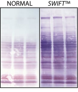
Apart from staining, there are additional products, such as Swift Transfer Pads, that can be used to reduce the necessary transfer times of proteins and prevent potential issues, like overheating. This will reduce the chances that the protein transfer could be incomplete. Swift Transfer Pads may also aid in transfers with particularly high molecular weights, though they can be used with any type of protein. Proteins that have high molecular weights may take longer to transfer and may have a higher probability of a poor transfer. Low molecular weight proteins will more easily pass through the pores of the membrane.
Labs may also use transfer buffers containing 20% methanol to further facilitate their transfers. If transfers are continually failing, there may be something wrong in the timing of the process itself.
Poor protein transfers are relatively easy to protect against and identify with the use of the right technology.G-Biosciences has a variety of products designed to provide better accuracy and speed throughout the process of protein transfer and western blotting. By ensuring proper protein transfer, many labs will be able drastically cut down on the time spent blotting and troubleshooting their blotting results.


