
Multiplexing your Western blots!!
Since its inception forty years ago, Western blotting has been a mainstay technique for molecular biologists around the world to detect and quantify proteins. Western blotting is a simple technique that involves the transfer of proteins (blotting) from labile polyacrylamide gels to resilient materials such as nitrocellulose or PVDF membranes using electrophoresis. Western blotting combines the resolving ability of SDS-PAGE and specificity of antibodies to detect the proteins adsorbed on the membrane. The detection methods such as chromogenic reactions or chemiluminescence allow detection and quantification of proteins. These methods are highly successful for routine applications in the lab but suffer from certain limitations also. For example, chemiluminescence is short-lived and requires quick processing of samples to obtain the best results.
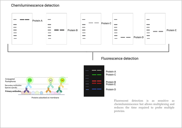
Another major shortcoming of existing methods is that it does not allow one to detect multiple proteins on the same blot at the same time. To detect different proteins on a blot, the membrane should be stripped and reprobed with different antibodies one by one. Sometimes more than one blot may be required to detect all the proteins under consideration, this becomes problematic when samples are limited. Moreover, stripping and reprobing can compromise the integrity of nitrocellulose membranes and can lead to the loss of adsorbed proteins, this can lead to serious errors in the quantification of proteins.
Schematic of fluorescent protein detection by Fluorescent western blotting
A major advancement in western blotting is the application of fluorescence in western blotting, this allows quantification of multiple target proteins in a single shot on a single blot. This also allows simplification of data analysis, because one can compare signals from different target protein bands as they all are on the same blot and therefore have been exposed to identical conditions of electrophoresis and transfer. Moreover, fluorescence is extremely sensitive and directly quantifiable as compared to chromogenic and chemiluminescence detection methods.
However, the transition from conventional western blotting to fluorescence western blotting requires special equipment equipped with fluorescence imaging channels that can excite different fluorophores and record various emission wavelengths. Simultaneous detection of multiple target proteins by fluorescence requires snapshot for each fluorescence signal and then combine these snapshots to produce a multiplex image.
There are certain important considerations to keep in mind as you switch to fluorescence Western blotting, they are mentioned below,
- Highly specific primary antibodies: Since fluorescence is very sensitive, primary antibodies should be extremely specific to the target protein and should not have cross-reactivity. Otherwise one can detect non-specific bands in fluorescence imaging. To detect different proteins, it is required to have species-specific antibodies against target proteins. For example, to detect four different proteins, one should use specific antibodies from rabbit, mice, rat and chicken. To detect actin protein one can use phalloidin conjugated to fluorescent tags.
- Secondary antibodies and spillover: Species-specific secondary antibodies tagged with highly fluorescent tags should be used. G-Biosciences provides a range of fluorescent tags HOOKTM (having a range of absorbance and emission spectra) that can be coupled to species-specific antibodies using NHS chemistry as mentioned in the previous blog article. It is very critical to choose fluorophores that don’t overlap significantly in their emission or absorbance spectrum to prevent spillover between imaging channels.
Note: one can also label primary antibodies with fluorophores but again the fluorophores used to label the antibodies should exhibit minimum spillover. - Nitrocellulose membranes: PVDF membranes can have some background fluorescence and therefore it is important to use nitrocellulose membrane that doesn’t give a background fluorescence.
- Blocking agent: it is recommended to use Casein (we recommend BlockTM Casein from G Biosciences) for blocking the blot.
- Optimising low abundance target detection: Protein targets that are low abundance should be detected using a brighter secondary fluorescent antibody.
Suggested readings
- Berkelman T. Fluorescent Western Blotting: High Sensitivity Detection of Multiple Targets. Curr Protoc Pharmacol. 2020;88(1):e72. DOI:10.1002/cpph.72
- Kondo Y, Higa S, Iwasaki T, et al. Sensitive detection of fluorescence in western blotting by merging images. PLoS One. 2018;13(1):e0191532. Published 2018 Jan 19. DOI:10.1371/journal.pone.0191532
- John M Walker, The protein protocols Handbook, 3rd Edition, Human Press
- Imaging Principles and Methods, GE healthcare Handbook


