Flow cytometry (FCM) is a multi-parameter cell analysis technique used for detecting and measuring the physical and chemical properties of a particular cell population, including cell count, cell size, and cell cycle. While the method has been routinely used for basic cell biological research, it also plays a vital role in the diagnosis, treatment, and management of certain bone and soft tissue tumors and blood cancers such as leukemia, lymphoma, and myeloma.
How Does It Work?
In this method, the sample (fluorescent-labeled cells derived from bone marrow, lymph nodes, or blood) is suspended in a fluid and injected into the flow cytometer instrument. A system of tubing, pipes, and valves forces the cells to pass through the instrument in a single file.
When the cells reach the interrogation point (or laser intercept), they pass through a focused laser beam or a similar light source, causing fluorescence emission and light scattering. Fluorescence intensity indicates the quantity of cellular constituents, while light scatter determines the size (forward angle scatter) and structure (right angle scatter) of individual cells.
The data is then collected and processed accordingly while the cell sample is properly disposed of in the waste container. In a cell sorter, however, the cell sample is passed on to a collection tube for post-experimental use.
Flow Cytometry and Cancer Research
Aside from its critical role in the field of molecular, plant, and marine biology, immunology, and virology, the importance of flow cytometry in cancer research cannot be underestimated.
While it was originally used in studying DNA content for cell ploidy and the determination of proliferation activity in human solid cancer types, high throughput, image-based FCM has taken the place of microscopy-based clinical tools as one of the most indispensable tools in cancer research.
And with the discovery of monoclonal antibodies and new fluorescent dyes, FCM gained an even wider application in the field of cancer research. At present, flow cytometry is used for:
- Cancer diagnosis. It is normally used to screen for abnormal DNA content which may indicate the presence of cancer.
- Specific histopathological diagnosis. It can detect RNA (for blood cancers) and specific tagged surface markers (for lymphoid and myeloid neoplasms).
- The presence of DNA aneuploidy and a high proportion of S-phase tumor cells may indicate tumor malignancy.
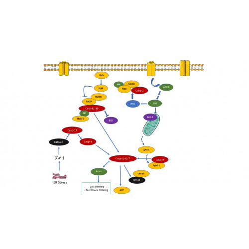
- Autophagy assessment. Autophagy is a process of cell death that occurs in normal cells and cells experiencing increased intracellular stress. Various mechanisms of autophagy are closely associated with promoting and suppressing the induction of tumor cells.
- Cancer treatment. Flow cytometry can be used to quantify the therapeutic and potential side effects of various cancer treatments. The identification of tumor antigens is also made possible by combining flow cytometry with monoclonal antibodies.
And here’s where it gets more exciting: Researchers have high hopes that this method will provide highly specific (“fingerprint-type”) information of a patient's tumor in the very near future. When this happens, determining the most appropriate and effective treatment can be made with greater accuracy and patient care can be improved dramatically.
Advantages of Flow Cytometry
There are several reasons why FCM has the approval of the scientific community, especially among those who are involved in cancer research.
Flow cytometry is extremely sensitive. FCM can detect the presence of a small number of cancer cells even if other testing methods provide no evidence of the disease. Since FCM is capable of identifying 1 in 10,000 (or even 70,000) cells, it can be particularly useful in monitoring remission and predicting the recurrence of the disease.
It provides quick turnaround time. FCM can analyze thousands of cells per second. This means patients can get their test results and bring them back to their clinician within hours. This is a definite game-changer, particularly in cases where prompt treatment is needed to ensure better outcomes.
Flow cytometry continues to evolve, and as the platform’s capabilities and precision keeps on improving, more effective and personalized cancer therapy can be expected in the horizon.


