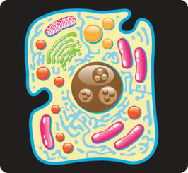
 Cells are made up of recognizable structures that each serve their own purposes and have their own characteristics. Common subcellular markers are often used to distinguish cells from each other and to identify potential irregularities within the cellular structure. Though there are a multitude of subcellular markers available, a few of them are more common and more easily identified than others.
Mitochondrial Markers
Mitochondria provide energy to the cell through Adenosine Triophosphate, abbreviated as ATP. Mitochondria have a variety of markers, as over 600 proteins have been discovered to be located within them. CSP1 (carbamoyl phosphate synthase 1) is commonly used in order to identify liver and kidney mitochondria. Prohibitin is a protein used throughout the process of mitochondrial respiration and can therefore also be used as a mitochondrial marker. Cytochrome c oxidase is a protein that is located within the inner membrane of mitochondria and which can serve as a good general purpose marker.
Golgi Markers
The Golgi apparatus is an organelle available in most cells which is designed to package vesicles and then direct them as needed. The position of the Golgi apparatus depends on the type of cell. In plants, the Golgi apparatus may be located throughout the cellular body. In mammals, the Golgi apparatus is generally positioned very close to the nucleus of the cell.
The Golgi apparatus may be difficult to separate from the trans-Golgi network, for which TGN38 staining is frequently used. TGN38 staining will stain the Golgi apparatus itself differently from the Golgi stack (also known as the cis Golgi network), thereby making it easier to view. The Golgi stack appears as a series of flattened disks with clusters emerging outward from it.
In addition to fulfilling the role of intracellular transportation, the Golgi apparatus may be able to synthesize carbohydrates and proteoglycans for use within the cell. Like many other organelles, the Golgi apparatus is dissolved and then reincorporated during cellular mitosis.
Nuclear Markers
There are three major nuclear structures that can be used as markers through the cellular body: chromatin, the nuclear envelope, and the nucleolus. Chromatin can be identified through dyes that bind DNA. These dyes will produce a chromatin marker. Two of the most common dyes are Hoechst 33258 and 33342. Certain proteins can also be used to target telomeres.
The nuclear envelope is the structure that encapsulates the nucleus within the cellular body. The nuclear envelope can generally be detected through the use of antibodies which detect Lamin A, Lamin B, and Lamin C. Finally, the nucleolus is the structure in the heart of the nucleus that is designed t construct RNA. Proteins available within the nucleoli an be used as marketers to detect the nucleolus.
Endosomal Markers
When molecules have to be transferred through the cell from the membrane to the individual organelles, they are encapsulated within endosomes. Endosomes are thus located throughout the cellular body. Proteins such as Rab5, Rab7, Rab9, and Rab11 are used by the endosomes to move molecules from the plasma membrane to endosomes. Consequently, they can be used as solid protein markers for the identification of endosomal structures. EEA1 is another similar protein that aids in the trafficking of molecules through endosomes. Clathrin and Adaptor protein-2 are more specific proteins which are able to mark certain vesicles which await the acceptance of transferred molecules.
Other types of subcellular marker include endoplasmic reticulum markers, cytoskeletal markers, and lysosomes. In different types of cells, certain markers may be more visible than others. Identifying subcellular markers often takes practice and patients, and individuals that are tasked with identifying subcellular markers may need to review them in a controlled laboratory setting before attempting to identify them in active experiments.


