
INTRODUCTION:
Many protein modification reagents for biotinylation and cross-linking involve reactive groups that conjugate through primary amines. Primary amines are found in all proteins and peptides as they make up the N-terminus and are also a component of the lysine residue side chains. Amines, lysine ε-amines and N-terminal α-amines, are the most abundant group in protein molecules and represent the most common target for biotinylation. For example, BSA contains 59 primary amines, of which up to 35 are available on the surface of the molecules and can be reacted with amine reactive esters.
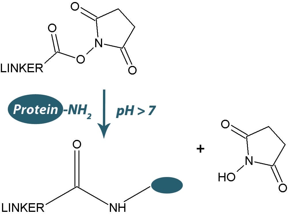
The most widely used amine reactive biotinylation and cross-linking reagents are the water insoluble N-hydroxysuccinimide (NHS) esters or the water soluble N-hydroxysulfosuccinimide (sulfo-NHS) esters.
The addition of a charged sulfonate (SO3-) on the N-hydroxysuccinimide ring of the sulfo-NHS esters results in their solubility in water (~10mM), but are not permeable to plasma membranes. The solubility and impermeability to plasma membranes makes them ideal for studying cell surface proteins as they will only react with the protein molecules on the outer surface of plasma membranes.
Both forms of the NHS esters hydrolyze rapidly in aqueous solutions (7 hours at pH7, minutes at pH9) and for this reason the NHS esters must be handled and stored appropriately. The NHS esters should be stored desiccated and by allowed to warm to ambient temperature before opening to avoid condensation. That being said, repeated opening and closing and inappropriate handling will lead to the introduction of moisture and hydrolysis of the NHS-esters.
Hydrolysis (and conjugation) results in the release of NHS that can be assayed with the following procedure.
Step 1: Materials Required
- NHS ester reagent
- DMSO or DMF for solubilization of NHS-esters
- Amine free buffer at pH7-8. We recommend a phosphate buffer, but avoid Tris and glycine buffers
- 0.5-1.0 NaOH
- Quartz cuvettes
- Spectrophotometer set at 260-280nm
Step 2: Straightforward Procedure
- Dissolve 1-2mg NHS ester reagent in 2ml amine free buffer. Prepare a control tube with 2ml amine free buffer.
NOTE: If using a non-water soluble reagent; first dissolve in 0.25ml DMSO or DMF and then add 2ml amine free buffer. Prepare a control tube in a similar fashion (0.25ml DMSO or DMF and 2ml amine free buffer) - Immediately, zero a spectrophotometer set at 260nm using the control tube and then measure the absorbance of the NHS-ester tube.
NOTE: If absorbance is >1.0, dilute entire solution with amine free buffer until readings are <1. - Add 100µl 0.5-1.0N NaOH to 1ml of the NHS ester from step 2. Vortex for 30 seconds
- Immediately (within 1 minute), measure the absorbance of the base hydrolyzed reagent.
Step 3: Simple Result Interpretation
- If the absorbance from step 4 is measurably greater than absorbance from step 2 then NHS reagent IS ACTIVE for amine reactive function
- If absorbance from step 4 is NOT measurably greater than absorbance from step 2 then NHS reagent IS HYDROLYZED and INACTIVE for amine reactive function. Discard and purchase new reagent
NHS Reactive Reagents
For a selection of amine reactive biotinylation and cross-linking reagents click on the handbooks below:


