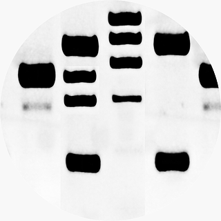
 Western blotting is an immunodetection technique used to first separate proteins by gel electrophoresis then to transfer those separated proteins from the gel matrix onto a secondary, more durable matrix, most commonly polyvinylidene difluoride (PVDF) or nitrocellulose.
After transferring to a membrane, the next step is blocking the membrane. This step is a crucial one in any Western blotting protocol, and failure to do so will result in antibodies binding to nonspecific binding sites on the membrane and causing a high background signal, which will make it very hard or impossible to detect the presence of the target protein. Nonspecific binding is essentially when an antibody doesn’t bind to its specific ligand, whether by sticking to the membrane, through the presence of proteins that bind to the Fc regions of the antibodies, or binding to the blocking buffer itself. Several different types of blocking buffers can be used to block a membrane; each with their own advantages and disadvantages. Which to choose depends largely on how the antibody or target protein will interact with the blocking buffer.
Non-Fat Dried Milk
- Typically used at 0.2-5% concentration
- Very cheap and widely available; stocked in almost any grocery store
- Contains phosphoproteins which can interfere with phosphospecific antibodies
Bovine Serum Albumin (BSA)
- Typically used at 0.2-5% concentration
- More expensive than non-fat dried milk, but still relatively inexpensive
- Can cross-react with phosphotyrosine antibodies
Purified Casein
- Typically used at 1% concentration
- Some of the cross-reactive components in non-fat dried milk are removed; however, it still does retain its ability to interfere with phosphospecific antibodies
Fish and Plant Derived Buffers
- Typically used at 2-10% concentration
- These typically do not cross react with animal antibodies
- Can interfere with protein-antibody interactions
Detergents
- Typically used at 0.01-0.1% concentration and frequently in conjunction with other blockers
- Most often Tween-20
- Useful in wash buffers to block the surface revealed when specifically bound proteins are physically removed during the washing process
- May disrupt protein-surface or protein-antibody binding
- Not permanent; can be washed away
Non-Protein Blockers
- Typically used at 0.5-2% concentration and often used in conjunction with other blockers
- Typically polyethylene glycol (PEG), polyvinylpyrrolidone (PVP), or polyvinyl alcohol (PVA)
- Readily binds to hydrophobic membranes and renders them non-binding and hydrophilic
Which blocking agent to use is more a process in optimization than a definite decision; the ideal choice should block non-specific binding to the membrane, block non-specific protein to protein interactions, not react to the primary or secondary antibody probes, minimize protein denaturation, and not interfere with protein to membrane bonds.
Resolving Blocking Issues
A typical problem with the blocking step when running a western blot is high background. This typically occurs because the blocking agent has not blocked the membrane effectively due to incomplete blocking or antibodies binding to proteins in the blocking buffer. Possible remedies for this problem include:
- Increase the concentration of the blocking buffer; too low a concentration and there may not be enough blocking agent to block all the non-specific binding sites
- Leave the blocking buffer on the membrane for a longer period of time to give it more time to bind to the non-specific binding sites
- Block the membrane at a higher temperature (up to at most room temperature)
- Change to a different type of blocking agent
Sometimes western blots can turn out with dark spots on it most commonly occurring because of the presence of clumps of blocking agents stuck onto the membrane. Solutions to this problem include:
- Ensure blocking agents are entirely dissolved before use
- Check blocking buffers for precipitate before use
- Filter the blocking buffer
- Add a detergent to minimize clumping of blocking agents
- Make fresh blocking buffer
Weak or no signal may be caused by blocking buffer interference with protein-antibody interaction:
- Reduce concentration of blocking buffer
- Detergents may interfere with protein-antibody interaction; repeat the assay without them present in the blocking buffer
- Change to a different type of blocking agent
Multiple non-specific bands (bands that aren’t the target protein) may appear due to insufficient blocking:
- Increase the blocking buffer concentration
- Block for a longer period of time
- Block at higher temperature
- Add Tween-20 as it can augment blocking efficiency
While blocking agents remove unwanted background they very often will interfere with protein-antibody binding. Finding the right combination and concentration of blocking agents can be a tricky process and very often leads to tradeoffs between low background and high sensitivity. The more blocking activity, the lower the background, but also the lower the signal. The less blocking activity, the higher the background, but also the higher the signal.


