
Question:
How do you perform protein electrophoresis?
The Protein Man Says:
After you have chosen the most appropriate method in studying your protein of interest, prepared your sample and identified the most suitable gel and buffer systems for your experiment, we now come to the best part – separating the protein of interest from the sample through electrophoresis and analyzing the results. How do you do it? Here's how.
 Performing the procedure. To provide optimum resolution and increase your chances of getting accurate results, you need to ensure that the temperature of the system is maintained at approximately 20oC during separation (this can be achieved with the help of a recirculating cooler). You also need to make sure that the appropriate running conditions are met.
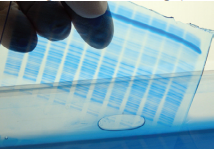
To do this, you need to prepare the electrophoresis cell, fill the inner and outer chambers with the right amounts of running buffer, heat your sample at 90 to 100oC for about five minutes and load the appropriate volume of the sample on the gel. Run the electrophoresis cell according to the manufacturer’s instructions, remove the gel from the cassette and use the appropriate stain to interpret your results.
Protein detection and analysis. Protein stains are usually used to analyze (both quantitatively and qualitatively) the protein band patterns produced during electrophoresis. However, since there are a lot of protein stains available for use (Coomassie blue/silver/fluorescent stains) you need to choose one that has a wide dynamic range, offers high sensitivity and reproducibility, compatible with downstream technologies and has a robust and fast protocol. You should likewise take the available imaging equipment into consideration as you choose an appropriate staining technique.
For best results, here are some guidelines you may want to consider when choosing an appropriate protein stain:
- Coomassie blue. Being the most popular anionic dye, Coomassie blue can stain almost all proteins with good linearity. It can also be used for mass spectrometry and protein identification.
- Silver stains. These dyes have the highest sensitivity but they unfortunately have a low dynamic range. In addition, they require complex and time-consuming protocols and do not offer sufficient reproducibility.
- Fluorescent stains. While fluorescent stains are ideal in almost all aspects, they are definitely more expensive as compared to the other two staining techniques. These staining dyes also require the use of a charged couple device (CCD) camera or fluorescence scanner for imaging. As such, their use is limited mostly to proteomics applications and 2D gels.
Keep these things in mind as you conduct your experiment and you will surely get the kind of results that you need.


