
Question:
What is the Process of Protein Separation by SDS-PAGE (PolyAcrylamide Gel Electrophoresis)?
The Protein Man Says:
Protein electrophoresis is a relatively simple, rapid and highly sensitive tool to study the properties of proteins. It is the principle tool in analytical chemistry, biochemistry, and molecular biology. The separation of proteins by electrophoresis is based on the fact that charged molecules will migrate through a matrix upon application of an electrical field. The matrix for protein electrophoresis separation is polyacrylamide.
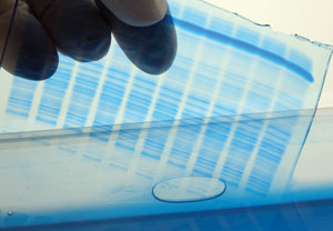
The chemical agents used to form polyacrylamide are monomeric acrylamide and N, N’-methylene-bis-acrylamide (bis-acrylamide). The most popular method for polymerizing acrylamide and bis-acrylamide is using TEMED (tetramethylethylenediamine) and ammonium persulfate. The polymerization of polyacrylamide creates a cross-linked network of tiny pores. This network of pores allows small protein molecules to pass through rapidly, while slowing the migration of larger proteins. This results in the separation of proteins based on their molecular size.
The size of pores in the polyacrylamide gel matrix is determined by the amount of total acrylamide used per unit volume and relative percentage of bis-acrylamide used. The effective range of polyacrylamide gel is between 3-30%.
SDS PAGE (SDS Polyacrylamide Gel Electrophoresis
Two fundamentally different types of gel system exist, non-dissociating (non-denaturing) and dissociating (denaturing). Non-dissociating (non-denaturing) system is designed to separate native protein under conditions that preserve protein function and activity. In contrast, a dissociating system is designed to denature protein into their constituent’s polypeptides and hence examines the polypeptide composition of samples. The denaturing system is probably the most common used and is known as SDS-PAGE.
The SDS used in an SDS-PAGE protein identification procedure stands for Sodium Dodecyl Sulfate, an anionic detergent that denatures the proteins and unfolds each polypeptide chain into a linear orientation. The SDS also applies an evenly distributed negative charge to each protein in accordance with its mass.
In SDS-PAGE, the protein mixture is denatured by heating at 100°C in the presence of excess SDS and a reducing reagent (β-mercaptoethanol, TCEP, DTT) is employed to break disulfide bonds. Under these conditions, all reduced polypeptide bind the same amount of SDS on a weight basis (1.4g SDS/g polypeptide) independent of the amino acid composition and sequence of the protein. The SDS-protein complex forms a rod with its length proportional to the molecular weight of the protein. All proteins are now negatively charged with similar charge density and thus can be separated on the basis of their size only.
SDS-PAGE is used mainly for the following purpose:
- Estimation of protein size.
- Determination of protein subunits or aggregation structures.
- Estimation of protein purity.
- Protein quantitation.
- Monitoring protein integrity.
- Comparison of the polypeptide composition of different samples.
- Analysis of the number and size of polypeptide subunits.
- Post-electrophoresis applications, such as Western blotting.


