We recently reviewed the advantages of total protein membrane stains as a loading control for Western blotting and demonstrated how they were an improvement on routinely used housekeeping genes.
What's best? Housekeeping genes or total protein for Western blot loading controls
One area that was not addressed is whether total protein stains can be used with the increasingly popular infrared (IR) imaging systems. There are several options for IR imaging systems on the market:
- Licor Odyssey
- Protein Simple FluorChem R System
- Azure Biosystems C500
- Syngene G:Box Chemi 66
- Vilber Fusion
These systems typically capture near infrared absorbance in 700 and 800nm channels, so the question is are there total protein stains that fluoresce at these absorbances. Secondly, one major advantage of the IR imaging systems can also act as a major drawback for total protein stains. The high level sensitivity of the IR imaging systems will result in the detection of residual membrane stains if not completely removed. So researchers will need a reversible protein stain with fluorescence in the near-IR wavelength.
G-Biosciences offers two total protein membrane stains:
1. Swift Membrane Stain
Swift Membrane Stain is a 30 second stain that stains total proteins on PVDF and nitrocellulose. Once stained and imaged, the stain is completely removed with a 30 second destain.
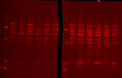
For IR Imaging systems the stain can be captured in the 700nm channel:
Following destaining with the supplied destaining solution, no residual signal is detected in the 700nm channel:
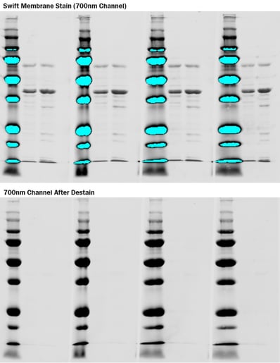
Pictures courtesy of Kristin Beede, University of Nebraska
2. Blot-FastStain
- Sensitivity: Transfer concentrates proteins on membranes and better exposes transferred protein to a membrane stain
- Durability: Proteins on membranes are more stable than in gel and therefore can be stored, reused and restained.
- Versatility: Nitrocellulose and PVDF membranes provide a better alternative for protein digestion for mass spec.
- Reversibility: Membranes are more amenable to reversible staining. Total protein profile can be visualized and quantitated prior to destaining; the membrane can then be processed in a conventional Western blot manner.
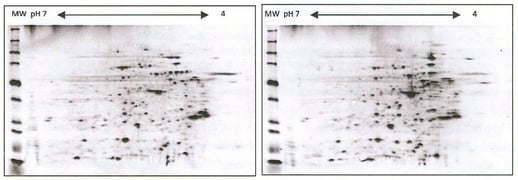
Figure: A431 cells transferred to nitrocellulose following 2D electrophoresis and stained with Blot-FastStain and imaged on the 800nm Odyssey channel.
In 2015, M.A. Collins et al utilized Blot-FastStain as an effective loading control for cerebrospinal fluid Western blots. They clearly demonstrated that Blot-FastStain was a reversible stain, with "no bands could be detected at the highest intensity setting of the Odyssey Clx imager".
Conclusion
G-Biosciences offers two reversible membrane stains that have independently been shown to be suitable for IR Imaging Systems. Swift Membrane Stain is visualized in the 700nm channel and Blot FastStain in the 800nm channel.